(ARMSTRONG, K AND JACKSON, S. (1972) Anatomy and Physiology for Nurses)
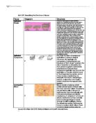
Connective Tissues
These tissues are responsible for cushioning, supporting and maintaining form within the body and are found all over the body linking and supporting organs. Connective tissue examples are; adipose (fatty tissues), loose connective tissue, dense fibrous connective tissue, elastic connective tissue, cartilage, osseous tissue (bone), tendons and ligaments.
I have chosen to look more at adipose (fatty tissue):
Adipose is a specialised type of connective tissue which stores lipids. It collects in large numbers and is shaped to be large round or oval cells. Adipose has a similar function to that of fibrous tissue, which throughout the body connects by irregular network of strands to form a cushion layer to support blood vessels, nerves and other organs.
Adipose is essential for insulation due to its low thermal capacity which allows the body to retain heat, thus functioning normally. Its other vital function is that of protection of delicate organs such as the eyes and kidneys. Fat cells offer this by forming liquid and being excellent at absorbing pressure as they cannot be flattened.
Adipose tissue is needed for the body to turn to in times of need due to it being able to form a food reserve. Excess carbohydrates are made into glycogen and turned into fat and stored within adipose tissue which when energy is required by the body converts back.
Adipose tissue is normally localised to certain depots within the body such as under the skin and around some organs like the kidney but may accumulate anywhere. In men, it usually represents 15-20% of body weight compared to 20-25% in women. Almost all adipose tissue in adults is known as white or brown adipose which is composed of tightly packed cells with a main feature being a large lipid droplet which is surrounded by a thin layer of cytoplasm bound by fine connective tissue fibres and blood vessels and its fibroblasts. Its also contains a flattened eccentric nucleus.
A recent finding in fat tissues is leptin which is useful for reducing appetite and is known to be made and released to travel through the blood, from fat cells.
( accessed 08 October 2004)
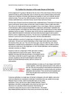
Muscular Tissue
Three types of muscular tissue are found in the body, these are; skeletal, smooth muscle and cardiac. This consists of cells which are capable of contraction as a result of nerve impulses, producing movement, both of the organism as a whole and of internal organs.
(HALE, W.G and MARGHAM, J.P and SAUNDERS, V.A. (1995) Biology Dictionary)
I have chosen to look more closely at cardiac muscle;
Cardiac muscle is specially striated muscle only found in the heart walls and has branching fibres. Cardiac muscle has striations but the fibres are linked laterally between the muscle fibres. Its contractions propel blood through blood vessels to every part of the body.
One centrally located nucleus is found per cell the nucleus is positioned centrally due to the nature of the contractions. Intercalated discs strengthen the fibres which enable contractions to occur and a bridge in between intercalated discs give strength and gap junctions will help to spread contractions. Its contractions are not under voluntary control. There are also many more mitochondria present in cardiac cells. It has to be able to contract without getting tired throughout the living life of the person or animal.
Microfibrils are linked in order for electric impulses to be passed from cell to cell to ensure contractions are maintained to be rapid, smooth and rhythmic. The contractile proteins causing the contractions are actin and myocin.
Bibliography
-
(ARMSTRONG, K. and JACKSON, S. 8th Edition, (1972) Anatomy and Physiology for Nurses, Baillier Tindall Publishers.)
-
(FREEMAN, W.H. and BRACEGIRDLE, B. 2nd Edition, (1967) An atlas of Histology, Heinemann Educational Books Ltd)
-
(HALE, W.G and MARGHAM, J.P and SAUNDERS, V.A. 2nd Edition, (1995) Collins Biology Dictionary.)
-
(Encarta accessed on 08 October 2004.)
-
(Histology Website, accesses 08 October 2004)