Circulatory System:
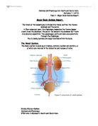
The main components of the circulatory system structure are; the human heart, arteries, arterioles, capillaries, venules, veins and the blood. The heart consists of 4 chambers which are each perfectly adapted to their function. The four chambers are called the left atria, right atria, left ventricle and the right ventricle. As you can see from figure 1. the left ventricular wall is significantly thicker than the right ventricular wall; it can be up to three times thicker. The left ventricle has a thicker muscular wall as it needs to be able to pump the blood out of the aorta with sufficient pressure to overcome the resistance of the systemic system. The walls of the atria chambers are made of very thin muscle; this is because they only need to be able to generate enough pressure to force the blood down the heart into the ventricles. (1,2)
Figure 1. (3)
The heart is in fact two pumps side by side. As you can see in figure 1. the right side of the heart receives the deoxygenated blood from the body and pumps it in the pulmonary system to the lung so it can be re-oxygenated. The left side of the heart receives the oxygenated blood from the lungs and transports it all around the body in the systemic system. (1,2)
The cardiac cycle consists of three phases; ventricular systole, diastole and atrial systole. The first stage of which is diastole, this means the heart muscle is relaxed. The internal volume in the heart increases causing blood to flow into the heart from the major veins. The blood firstly flows into the atria, as the atria fill with blood the blood pushes the atrioventricular valves open allowing blood to flow down into the ventricles. The second stage of the process is called atrial systole. This means the atria is contracting. Both the right and left atrium contract together. This contraction increases the pressure slightly which forces the blood down into the ventricles. As the blood enters the ventricles it causes the walls to expand to ensure they are full of as much blood as possible. The final stage Ventricular systole, when the ventricles are full of blood they start to contract. As the ventricles start to contract the pressure rises above the atria causing the atrioventricular valves to snap shut to prevent back flow into the atria. The valves can only open one way, they are prevented from going ‘inside out’ by the tendinous cords attaching them to the wall of the ventricles. These cords are called chordate tendinae. There is now a short time in which all four valves are closed. The pressure rises very quickly in the ventricles. The contraction starts in the Apex of the heart which allows the blood to be pushed up and out of the ventricles towards the arteries. The semilunar valves open and the blood leaves the heart. The contraction lasts a very short amount of time, and then the ventricle walls relax causing the heart to start refilling. The muscle of the heart is a special kind of muscle called cardiac muscle which is myogenic, this means it doesn’t need any stimulation from nerves to make it contract. It is able to initiates it own contractions. The heart is able to continue to contract rhythmically even if it is not connected to the body. The heart muscle can result in inefficient pumping called fibrillation if the contraction of the heart chambers are not synchronised. To coordinate the contractions the heart has its own mechanism called the sinoatrial node (SAN) which acts as the hearts pacemaker. It is a small patch of tissue located in the top right atrium wall. The tissue sends out waves of electrical excitation at regular intervals to initiate contractions. This wave of excitation spreads quickly through the atrial walls by travelling through the membranes of the muscle tissue. As the wave of excitation passes it causes the muscle cells to contract, this is atrial systole. At the base of the atria is ring of fibrous tissue which does not allow the electrical impulses to pass straight through this. The only way the electrical impulse can travel from the atria to the ventricles is via a group of specialised muscle cells called the atrio-ventricular node (AVN) which is at the top of the inter-ventricular septum. When the wave of excitation reaches this point there is a slight delay which gives the atria time to fully empty. After this slight delay the wave of excitation is carried away from the AV node and down the specialised conducting tissue called purkyne tissue. The purkyne tissue runs down the ventricular septum to the base of the heart which is called the Apex. Also in the septum is muscle fibres called the bundle of his, these fibres divides into right and left branches at the base of the ventricles. They spread through muscular walls in purkyne tissue. As the wave of excitation spreads upwards from the Apex of the ventricles the muscle starts to contract. This means the ventricles contract from the base upwards pushing the blood up and out to the major arteries located at the top of the heart. This is followed by a short amount of time where there are no impulses passing through the heart muscle, this is the heart relaxing and diastole occurring. (1,2)
(4)
(5)
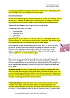
The function of arteries is to carry blood away from the heart. As the blood is just leaving the heart it is at very high pressure so the arteries need to be able to withstand this. Arteries are adapted to their function in multiple ways; their lumen is relatively small to maintain high pressure in the blood, the wall is relatively thick and contains collagen which is a fibrous protein to give it the strength it will need to with stand the high pressure, elastic fibres in the wall allow it to stretch and recoil when the heart pumps, the recoiling helps maintain high pressure while the heart relaxes, the smooth muscle in the wall can contract and constrict the artery, constriction narrows the lumen and the endothelium can be unfolded when the artery stretches. In the circulatory system there are five different types of vessels, they include; arteries, arterioles, capillaries, venules and veins. Each of the vessels are highly adapted to their function, capillaries are tiny blood vessels which are approximately 7-10 μm wide. They are the same width as a red blood cell so the cells are able to pass along the capillary but they do have to be squeezed through. This slows the flow of the red blood cells down as they can only pass through one at a time allowing efficient exchange between blood and the tissues. The structure of a capillary is a single layer of flattened endothelium cells. Because the walls are so thin it increases their overall surface area making gaseous exchange fast and efficient. In the capillary wall are tiny pores which allow other substances to be exchanged between the capillaries and tissues including phagocytes to migrate into tissues. Capillaries are found all over the body, another role of a capillary is to oxygenate deoxygenated blood. Because the capillaries are so small and thin they are able to get close enough to the alveoli in the lungs so red blood cells can be oxygenated. Arteries are the extreme reverse of capillaries. Arteries have thick walls which are made up from smooth muscle and elastic tissue. The walls need to be thick and strong so they are able to with stand the high blood pressure without bursting or leaking. As blood is forced through the artery with every heart beat the artery walls are forced to bulge, the elastic fibre layer allows the wall to stretch and recoil back to its original shape. This process is known as elastic recoil. Because of this the high blood pressure is maintained and the flow is kept smooth. The lumen, the space blood flows through, is relatively small keeping the blood under immense pressure. Like a capillary and a vein an arteries inner layer is made of flattened endothelium cells, this is a very smooth layer which helps the blood to flow with as little friction as possible. The final outer layer is made out of collagen fibres. This tough fibrous layer protecting the artery from damage when we move around. Veins have much thinner walls than arteries. This is because the veins are working with blood at a lot lower pressure. A vein has a wider lumen than an artery; this is to keep the blood travelling at a slower pace. Like an artery a veins outer wall is made out of collagen fibres to protect it as we move around. Its inner layer, the endothelial wall, is there to reduce friction allowing blood to run smoothly without disruption. The main difference between an artery and a vein is that veins contain valves. Vales are put in place to prevent back flow and to keep the blood moving in the right direction, this is also their function in the heart. Because the blood is at such a low pressure it could easily start to flow in the wrong direction. The blood is able to keep flowing back to the heart through the veins because our skeletal muscles contract which squeezes the veins. This increases the pressure inside the veins forcing the valves behind to shut and the valve in front to open allowing blood to flow through. Arterioles have a thin wall mainly consisting of muscle fibres, but also contain some elastic fibres. When the muscle fibres in the wall contract this decreases the size of the lumen. When the muscle fibres relax the lumen is widened. Arterioles are able to increase or decrease blood flow to particular tissues at any point in time. They are also used as a way of regulating the blood pressure. The walls of venules are very thin and consist of muscle fibres and elastic tissue. They are just like small veins; their function is to carry blood from capillaries back to the veins. (1,2)
(6)
Valves in a vein (7)
Arterioles and Venules (8)
There are 5 main components that make up the blood in a human body, these are; erythrocytes (red blood cells), white blood cells (leucocytes), platelets, plasma and Immunoglobulins. The function of the red blood cells is to carry oxygen and distribute it to respiring cells and tissues; they also carry waste products such as carbon dioxide to the lungs. White blood cells are another of the main components, they are a vital part of the body’s defence system, and they fight infection in ways including phagocytosis. There are four types of white blood cells; neutrophils, lymphocytes (B&T) and monocytes. Each of these types of white blood cells have different functions. Neutrophils have small granules in its cytoplasm; they engulf microorganisms or other invading particles by phagocytosis. Lymphocytes B&T both have a darkly stained nucleus surrounded by a thin layer of cytoplasm. Both of these white blood cells are cells of the immune system but have different functions, B Lymphocytes produce anti-bodies and T Lymphocytes have several functions including cells destruction. Lastly monocytes, the largest kind of leucocyte have a large lobed nucleus and clear cytoplasm.(2) These cells spend up to 3 days inside the circulatory system before travelling into tissues where they become macrophages. As macrophages they engulf microorganisms and other foreign materials. Platelets are tiny fragments that are formed in the bone marrow. These fragments are crucial in the blood clotting process.(9) Plasma is a yellow coloured fluid that serves as a transport system carrying and delivering various materials between the cells. 92% of plasma consists of water, but it also contains other components such as salts, proteins, lipids and glucose. Blood plasma makes up 55% of the total volume of blood in a human body. Plasma also insures blood cells can flow throughout the body to where they are needed. (10) Lastly, Immunoglobulins are protective antibodies that are generated by white blood cells. These form when recovering from an infection or receiving an immunisation. Once formed these antibodies protect an individual from a future attack of the infection. (9)
Respiratory System:
The main structure of the respiratory system consists of; mouth and nose, trachea, bronchi, lungs and the alveoli. Additionally the ribs, intercostals muscles and the diaphragm are heavily involved in ventilation. The most important function of the system is to get oxygen transported to respiring tissues and cells and remove carbon dioxide. This overall process is aided by several smaller processes that include; gaseous exchange, external respiration and cellular respiration. (11) Respiration can anaerobically or aerobically. Lungs are a pair of inflatable structures lying within the chest cavity. The pathway for oxygen to reach the lungs is through the nose or mouth, along the trachea, bronchi and bronchioles. Each of these parts of the airway are specially adapted to their function. The trachea has ‘C’ shape rings around it which helps the airway to stay open when we breathe in and out. The lining of the trachea is made of ciliated epithelium cells and goblet cells. (2) The actual site of the gas exchange is the alveoli, they are tiny hollow sacs made of squamous epithelium cells. The walls of the alveoli are where gaseous exchange takes place. Gases can pass through both ways of the alveoli, oxygen passes from the air sacs to the blood in the capillaries and carbon dioxide passes from the capillaries to the alveoli. The lungs are protected by ribs. The action of the ribs and diaphragm, a layer of muscular tissue located underneath the lungs, help to produce breathing movements. Lungs are primarily adapted to their function with many specialised features for efficient gas exchange. These are; a large surface area, a barrier permeable to oxygen and carbon dioxide, thin barrier to reduce diffusion distance and maintaining the diffusion gradient. The larger the surface area the more space there is for molecules to pass through it. On their own alveoli are very small but as there are numerous alveoli in our lungs the total surface area has been calculated to be 70m2 overall. The membrane which is permeable to oxygen and carbon dioxide is the same membrane that surrounds the outside of cells. The walls are very thin to allow diffusion of oxygen and carbon dioxide to occur readily. Adaptations that have been put in place for rapid diffusion are; the wall of the alveoli and the capillary are very thin (approximately one cell thick), this means that oxygen molecules only have to diffuse the distance of two cells thick. Apart from the structures being only one cell thick they are also made of squamous cells which are flattened cells. As you can see from figure 2 the alveolus wall and capillary are in close proximity to each other, again this makes for gas exchange to be efficient. In capillaries the rate of the red blood cells flowing through them is greatly reduced as the capillaries are so narrow. Because they are so narrow the erythrocytes are squeezed against the capillary wall making them close to the air within the alveoli. The lungs must produce a substance called surfactant in order to work properly. Without surfactant the alveolus would collapse due to cohesive forces between the lining of the air sac and the water molecules. Surfactant also lowers the surface tension of the alveoli reducing the amount of effort needed to breathe in and inflate the lungs. Lastly, in order to have diffusion occurring rapidly a steep diffusion gradient needs to be maintained. This means that there needs to be a high concentration of the ‘supply side’ and a low concentration on the ‘demand side’. To make this happen there needs to be a fresh supply of molecules on one side and molecules need to be removed from the other side. This is achieved by ventilation and the blood transport system. Blood is the transport route of carbon dioxide from tissues and cells. Inhaling and exhaling fills the alveoli with fresh air, in addition to this ventilation removes the carbon dioxide that has diffused into the alveoli. This results in there being a low concentration of carbon dioxide in the lungs at anytime and a consistently high concentration of oxygen. (1)
Figure 2 (12)
Respiration happens due to inspiration and expiration. During inhalation the diaphragm becomes flattened causing digestive organs to be pushed down. The external intercostals muscle then contracts forcing the ribs to rise which then increases the overall volume of the chest cavity. This causes the pressure within the chest cavity to drop lower than the atmospheric pressure. Air is forced to flow into the lungs. During exhaling the reverse processes happen, the diaphragm relaxes as it pushes back into alignment by the displaced digestive organs that are underneath it. The volume of the chest cavity decreases as the external intercostals muscles relax. The pressure on the lungs increases above the atmospheric pressure and the air moves out of the lungs. (1)
(12)
Nervous System:
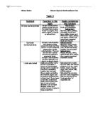
The nervous system allows every living organism to respond to their environment, the nervous system uses receptors and effectors to help aid it in responding adaptively to internal and external environments. This is a continuous and complex process involving coordination from different areas of the body including muscles, hormones and nerves. The nervous system is broken down into many different sub sections; the second division consists of the Central Nervous System and the Peripheral Nervous System. The central nervous system is made up on the human brain and the spinal cord. The central nervous system is concerned with all life functions and psychological processes. The spinal cord receives and transmits information to and from the brain, however the main maintains life. It is involved in higher functions and psychological processes. The peripheral system is made up of the receptors and effectors; these are the nerves that run from the central nervous system to the rest of the human body and back to the central nervous system. The peripheral system breaks down into two components; these are the somatic nervous system and the autonomic nervous system. Somatic system involves the sensory and the motor neurones which run the skeletal muscles. The somatic nervous system links the parts of the central nervous system and the peripheral nervous system together to help the human body deal with sensation, perception and other cognitive processes the human body may encounter. It also transmits information to and from the sensors and to and from the central nervous system. This system produces voluntary behaviour. The autonomic nervous system only consists of motor neurones which are responsible for carrying impulses to the effectors rather than the skeletal muscles. It transmits information to and from internal organs to sustain life processes. This system meditates involuntary control and responses in the functioning of the body systems which happen automatically. The autonomic nervous system controls processes such as heart rate and digestion. The part of the body that controls the autonomic nervous system is a structure in the brain called the hypothalamus. The actions of the ANS are always involuntary, it is able to adjust our breathing and changes in heart rate when it is necessary e.g. when we participate in exercise and digestion with no contribution. In some cases we are able to control some of these processes ourselves as we can hold our breath or we can raise our heart rate by exercising. Our autonomic nervous system is working every minute of every day of our lives unlike the somatic system which shuts down while we are sleeping. The last division of the nervous system which again consists of two components is the Sympathetic Nervous System and the Parasympathetic Nervous System. These two systems use different neurotransmitters so often have antagonistic effects. The sympathetic nervous system generally increases bodily activity where as parasympathetic nervous system maintains or decreases bodily activities. (13) (14) both these nervous systems have opposite effects on the functioning of the body but balance out.
(15)
References:
(1) AS Biology, OCR, Pete Kennedy and Frank Sochacki, Series Editor: Sue Hocking, Published 2008, Pages: 46-47, 54-61
(2) AS/A2 Human Biology, OCR, Barbara Geatrell, Pauline Lowrie and Alan Tiley. Series Editor: Fran Fuller. Published 2008, Pages: 6, 36-38, 42-43, 48-51
(3)
(4)
(5)
(6)
(7)
(8)
(9)
(10)
(11) Health and Social Care A2, AQA, by Richard Smithson, Published 2006, Chapter 9, Pages; 281-283, 285-288
(12)
(13) A2 Revise Biology, OCR, Jennifer Gregory, Lanto Stevens and Richard Fosbery, Series Editor: Sue Hocking, Published 2001, Pages: 84
(14) Daisy’s psychology pack
(15)
(16)
(17)
(18)