“Synarthroses are fixed joints at which there is no movement. The articular surfaces are joined by tough fibrous tissues. Often the edges of the bones are dovetailed into one another as in the sutures of the skull.”
(H.G.Q. Rowett 1999)
Cartilaginous joints
Also called Amphiarthroses joints.
These are joints joined by tough fibrous cartilage which provides stability and shock absorption. There is small amount of movement in these joints. For example, the articulations between the lumbar bones due to intervertebral discs of cartilage.
“Aphiarthroses are joints at which slight movement is possible. A pad of cartilage lies between the bone surfaces, and there is a fibrous capsule to hold the bones and cartilage in place.”
(H.G.Q. Rowett 1999)
Synovial joints
Also called Diarthroses joints.
These are the most common joints in the body. They allow the widest range of movement and are important in terms of physical activity.
“Synovial joints are known as freely moveable joints, though at some of them the movement is restricted by the shape of the articulating surfaces and by the ligaments which hold the bones together.”
(H.G.Q. Rowett 1999)
There are six types of synovial joints.
- Hinge
- Gliding
- Pivot
- Saddle
- Condyloid
Ball and Socket
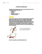
This is where a round end of one bone fits into a cup shape cavity of another bone. This joint allows movement in many different directions,
eg. this joint is at the hip and the shoulder.
Movement in this joint is- flexion, extension, shoulder & hip flexion and extension, abduction, adduction and circumduction.
Hinge
This joint is like a door hinge. It can swing open until it’s straight,
E.g., the elbow joint and knee joint.
Movement in this joint is- flexion, extension, plantar flexion and dorsi flexion.
Pivot
This is where a ring on one bone fits over a peg on the other allowing rotation, e.g. the neck joint between the atlas and axis.
This joint can have rotational movement, flexion and extension.
Saddle
The ends of the joints are shaped like saddles and fits snugly together.
E.g. joint at the base of the thumb.
Condyloid
A bump on one bone sits in the hollow formed by another bone or bones. Movement is back and forward and from side to side, e.g. the wrist joint.
Movement in this joint is- plantar flexion and Dorsi flexion.
These joints allow different types of movement.
Flexion- the angle of the joint is decreased.
Extension- the angle of the joint is increased.
Shoulder & hip flexion- limb moves forward, flexion to the front of the body.
Shoulder & hip extension- limb moves backward of the body.
Plantar flexion- toes pointing down, angle of the joint increased.
Dorsi flexion- toes pointing up, angle of the joint is decreased.
Hyperextension- the angle of the joint is more than 180 degrees, i.e. bending backwards.
Abduction- movement away from the midline of the body.
Adduction- movement towards the midline of the body.
Circumduction- a circular pattern, the limb moves in a curcular manor ad includes flexion, extension, abduction and adduction.
(Task 3)
A world class gymnast who competes and trains regularly is likely to suffer from both joint and bone homeostatic imbalances at some point in their life.
One example of a bone homeostatic imbalance is Osteoporosis. This is a disorder characterised by decreased bone mass, owing to loss of bone mineral, and increase susceptibility to fractures. A gymnast may be susceptible to Osteoporosis as she has smaller bones and has less adipose tissue, which is a great source of estrogen. Estrogen is a female hormone, which protects against bone mineral loss during young adulthood. Also adipose tissue is a good source of Estrone.
There are causes which can lead to Osteoporosis. Dieting can lead to a decreased bone mineral content as the body draws calcium from bone to make up for the calcium missing from the diet.
Excess protein can lead to loss of bone mineral as doubling protein intake in diets increases urinary calcium excretion by 50%.
A high fibre diet may decrease calcium absorption from the intestine, since dietary fibre may bind with calcium.
Eating disorders or excessive exercise causes menstrual irregularity. This can also lead to a decrease in bone mineral content.
Having severe Osteoporosis can create a physical feature, Dowager’s Hump. This is when so much bone has been lost the spinal cord loses strength and individual vertebrae collapse because of what are known as crush fractures. The spine develops a hunchback, which is very painful and the person has loss of height.
A person needs to have a good diet. Calcium can help to prevent or treat osteoporosis. Milk is fortified with calcium.
Vitamins A C & D are necessary in the diet for optimal bone development. Vitamin C is essential for collagen production, vitamin D improves efficiency of intestinal absorption of calcium and vitamin A aids in bone development.
Weight bearing activity helps to maintain bone density during adolescence and young adulthood as it applies mechanical stress. A weight bearing activity refers to activity in which the skeletal system must support the body weight i.e. walking, hiking and cross country skiing.
Another example of a bone homeostatic imbalance is a Fracture. A fracture to the tibia or fibula would cause great pain to the athlete.
“A twisting force to a stationary foot or direct violence cause a fracture of both.”
(David S. Muckel 1977)
Fractures of the fibula cause tenderness over bone but they can be treated by a bandage or strapping for 3 weeks if they do not involve the stability of the ankle joint.
Prolonged road work and repeated minor trauma to the shaft of the bone can cause a stress fracture. The athlete would have a dull aching pain over the bone. It would take 4 weeks for an X-ray to indicate the fracture by the periosteal reaction. To treat this the athlete would have to rest for 3-4 weeks, then reduce their activity, wearing protective footwear such as thick soled shoes.
Diagram of fractures of Tibia and Fibula
One example of a joint homeostatic imbalance that a gymnast may suffer from is ‘Tennis Elbow’. Tennis elbow or Lateral Epicondylitis is a condition when the outer part of the elbow is very tender and painful. Tennis elbow is a very similar condition to Golfers Elbow but it affects the other side of the elbow. Having this condition is very painful. Movement of the elbow and also movement involving lifting with the hand on top is painful.
Lateral epicondylitus is common in people who overuse their arm, not necessarily in tennis. A direct injury, bang or strain can sometimes cause the inflammation. Sometimes the muscles are actually partially torn.
“Sometimes the problem is partially or completely due to a neck problem, which is causing pain in the elbow via the nerves from the neck.”
(yahoo.com 2000)
There are a number of things that can treat this joint homeostatic imbalance.
- “Rest helps, with avoidance of the activities which overuse the elbow.
- Physiotherapy treatments, which may include heat or ultrasound therapy.
- Use of anti-inflammatory drugs and ordinary painkillers.
- The doctor may suggest an injection of a small dose of steroid to the affected area. This is not the sort that is banned from athletes. If used it can last for up to three months and although it may need to be repeated you seldom need more than two or possibly three injections.
- You can buy a brace from a sport shop or pharmaceutical supplier, which can be helpful. This is probably largely because it reduces the amount you can use your elbow.”
(Yahoo.com 2000)
Diagram of Elbow Joint
Another example of a joint homeostatic imbalance is ‘Meniscal Damage’ which is an injury to the knee. The two menisci are often called ‘cartilage’s’ since they consist of fibro-cartilage. They are commonly damaged during sporting activities. Treatment for ligamentous weakness is for it to be removed or later osteoarthritis may develop.
Cause of Meniscal Damage- “A menisci is torn by a rotating force carried out when the joint is partially flexed. This rotating force ‘sucks’ the meniscus concerned towards the centre of the joint when it I crushed between the extending tibia and femur.”
(David S. Muckle 1977)
“If a portion of cartilage becomes caught in the inter-condylar notch then locking follows.”
(David S. Muckle 1977)
“Vigorous internal rotation of the femur on the tibia with the knee flexed and weight bearing traps an excessively mobile medial meniscus, and extension tears it.”
(David S. Muckle 1977)
After this happens the knee may lock, sometimes an effusion with snapping or locking may be the sole complaint. The knee may give way and tenderness around the joint may be detected.
To treat this for the first locking episode - rest, traction and physiotherapy is needed for about 2-3 weeks. If the damage is recurrent then surgical excision may be needed, as tears of the body never heal. The athlete can return to sports activity in 6-8 weeks. If they have postoperative rest for 10 days with static exercises, then physiotherapy.
Bibliography
Anatomy & Physiology, forth edition. H.G.Q. Rowett 1999
John Murry (Publishers) Ltd. London.
Sports Injuries, David S. Muckel 1997
Published by Orial Press limited, Northumberland.
Other sources
Notes from class, Jenny Brown 2000.
2000
.
Gliding Joint
This is flat surfaces which can glide over each other, with little movement
in all directions, e.g. joints between carples, and between tarels.