The synovial joints have six sub-classifications. The first of these sub-classifications is the ball and socket joint; this joint provides the highest range of movement and is found at the shoulders and hips. These movements include; flexion, extension, rotation, adduction, abduction and circumbduction. The range of movement found at this joint can be seen fully during a match of tennis as the ball and socket joint of the shoulder; adducts, abducts and rotates throughout the match.
The second of these six joints is the hinge joint. The hinge joint can be found at the elbows, knees, fingers and toes. Its range of movement is limited to flexion and extension. The hinge joints full range of movement can be seen when a 100 metre sprinter is running, as the conventional runner keeps the elbows in a flexed position while the knee flexes and extends to cover the distance in the fastest possible time.
The third of the six joints is the pivot joint. The pivot joint can be found I the neck at the atlas and axis and between the ulna and radius. The pivot joint allows the ulna and radius to perform types of movement known as supination and pronation while, it allows the head to rotate to a certain extent atop of the atlas and axis. These movements can be seen during a table tennis match when, a person is serving or returning a shot while applying spin to the ball through the pivoting action of the ulna and radius.
The fourth of the six joints is the condyloid joint. The condyloid joint can be found in the wrists and ankles, it allows flexion and extension but, rotation is limited. The movements available at this joint can be seen when a basketball player is releasing a jump shot and following through with their fingertips.
The fifth of the six joints is the saddle joint. The saddle joint can be found in the thumb joint, it allows flexion, extension, adduction, abduction and circumbduction. The movements available at this joint can be seen during, a cycling race where the saddle joint flexes to allow the rider to grip the handlebars correctly to maintain balance.
The last of the six joints is the sliding joint. The sliding joint can be found in the wrists and ankles, more specifically the intercarpal and intertarsal joints. The joint does not perform a specific form of movement but, it does allow the carpals of the wrist and ankle to glide/slide over each other.
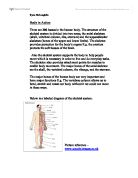
The Skeleton and how it responds to Exercise
Short and Long Term
The skeleton develops through weight bearing activities. These developments affect not only the bones of the skeleton but also, the cartilage, the ligaments and the tendons.
Short term responses to exercise for the skeleton include; the synovial fluid in the synovial joints becoming less viscous; the synovial membrane producing more synovial fluid to cope with the task at hand, in turn, reducing the amount of friction between the articular cartilage of the two bones forming the joint and, a greater range of movement is available at a joint, this is because of the ligaments and tendons in the joint becoming warm.
Long term responses to exercises include; greater bone density due to; larger amounts of calcium and collagen being released into bone, the ligaments around a joint become stronger and more elastic due to repeated use, this in turn increases the range available at a joint and, the articular cartilage of the bones becomes thicker to deal with the higher levels of mechanical stress being applied to the joints this will help later in life as the chances of suffering from arthritis are lowered.
The above are all positive factors in that they help to prevent osteoporosis as there is a higher level of calcium in the bone before the calcium begins to be lost as a person ages. Secondly, the levels of collagen in the bones reduce the chances of them becoming brittle with age as the body produces less protein as we age and this can sometimes lead to alterations in the makeup of the our bone.
There are possible negative effects, these include; the snapping of ligaments and tendons due to them being worked to much; instead of becoming more flexible and elastic they become hard and brittle, hyperextension of a joint, a lack of synovial fluid which will lead to the articular cartilage rubbing against each other with a higher amount of friction, in later life this could lead to arthritis, another effect due to a lack of synovial fluid is infection in the joint and, if there is no calcium then the bones will not become more dense, leaving them brittle and more prone to breaks and fractures.
Thickened layers of hyaline cartilage would allow the body to cope with larger amounts of shock and mechanical stress, which if applied to a thinner layer of cartilage could lead to trauma of a bone; in the form of a compact/compound fracture or a hairline fracture.
These thickened layers of cartilage help to cushion the vertebrae, improve posture and reduce the pressure on the lumbar of the spine; which bears most of the body’s weight. This reduced amount of stress in the lower back will benefit many peoples’ not just athletes’ lives, as they will be more mobile in older age and less likely to develop posture problems.
Also the facet joints between each vertebra are covered in articular cartilage making turning and bending forwards and backwards a smoother action as the cartilage reduces friction and stops the bones from grinding away at each other. In the long run the thicker the layers of cartilage between the facet joints, the longer the amount of time a person will go without lower back pain.