Joints
There are three types of joints these are:
- Fibrous
- Cartilaginous
- Synovial
Fibrous or Fixed Joint-
A fibrous or fixed joint has no movement at all. Tough fibrous tissue lies between the ends of the bone, which are dove tailed together. For example the sutures between the bones in the skull.
Cartilaginous Joint (slightly moveable)-
A cartilaginous joint allows some slight movement. The ends of bones are covered in articular or hyaline cartilage, separated by pads of white fibrocartilage. Slight Movement is made possible because the pads of cartilage compress. In addition these pads act as shock absorbers. For example the intervertabal discs in the spine.
Synovial Joint (freely moving)-
A synovial joint is a freely moving joint and is characterized by the presence of joint capsule and cavity. The type of joint is subdivided according to movement possibilities, which are dictated by the bony surfaces that form the joint. For example cartilage that reduce fiction in the knee joint.
There are different types of synovial joints that allow for different types of movement, they include:
- Ball and Socket
- Pivot
- Condyloid
- Hinge
- Saddle
Ball and Socket Joint-
The almost hemispherical surface of one bone fits into a cup like depression of the other. Movement is free in every direction. To facilitate movement, such joints are rimmed with cartilage and lubricated by synovial fluid. The bones are kept in place by ligaments and moved by muscles. For example the hip joint is a very strong ball and socket joint which can carry a load many times in excess of the normal body weight, in sport an example of this would be a strike in football.
Pivot Joint-
The ring-like atlas vertebra fits over a peg on the axis vertebra allowing the head to rotate. A bony ring rotates round the pivot (axis) of another bone. For example the pivot joint in your neck allows you to turn your head from side to side looking for team mates or opponents in sports.
Condyloid Joint-
Movements in two places is possible in this joint this is due to the dome shape surface of one bone fits into the hollow formed by one or more other bones forming the joint. Rotation is prevented by the attachment of ligaments. It is similar to the ball and socket joint, the joint allows circular motion. In the condyloid joint, the ball rests up against the end of a bone rather than inside a socket. The carpals of the wrist rest against the end of the radius bone of the forearm. For example in cricket the condyloid joint helps the bowler produce the perfect bowl.
Hinge Joint-
Movement is allowed in one place this is only due to the shape of the bones as well as the strong ligaments which prevent side to side movement, the
Joint allows both flexion and extension. The joint acts like a hinge on a door, allowing the joint to “open” and “close.” For example the hinge joint at the elbow will allow you to do a bicep curl with weights.
Saddle Joint-
Convex and concave bone surfaces are placed against each other. This allows movement in two places at right angles to each other. Where the thumb meets the wrist, the bones fit up against each other like a saddle fits over the back of a horse. For example any racket sport would show an example of this joint.
Synovial Joint Structure
The synovial joint is made up of a number of elements:
-
Articular or hyaline cartilage: a smooth, shiny cartilage that covers the ends of bones and absorbs synovial fluid.
-
Joint Capsule: a sleeve of fibrous tissue surrounding the joint.
-
Ligament: a sleeve of tough, fibrous connective tissue, which is an extension of the joint capsule.
-
Synovial Membrane: a sheet of epithelial cells inside the joint capsule
-
Synovial Fluid: the fluid enclosed in a joint, some of which is absorbed by hyaline cartilage during exercise.
-
Pad of Fat: pads of fat that occupy gaps in and around the joint.
-
Bursae: are little sacs of synovial fluid.
-
Menisci: are layers of fibro-cartilage located at the articulating surfaces of joints.
Here is an example of the components what make up the knee joint:
Movement at the Synovial Joints
Lots of movement takes place at the Synovial joints these include:
- Flexion
- Extension
- Abduction
- Adduction
- Circumduction
- Rotation
- Supination
- Pronation
- Eversion
- Inversion
- Dorsiflexion
- Plantarflexion
Flexion
Flexion is the bending, decreasing the angle between two bones. Flexion occurs at the trunk when bending forwards or sideways, at the shoulder when moving the arms or shoulders forward, in the arm when bending the elbow, in the hip when the thigh moves forwards and at the knee when bending the leg. Examples of this would be at the elbow when doing a bicep curl or at the knee during the phases of striking a ball.
Extension
Extension is the opposite of flexion, as it is increasing the angle between two bones. Extension occurs when bending backwards at the trunk, the shoulder movements of the arm or bringing the shoulder backwards, when the arm straightens at the elbow, at the hip when the thigh moves backwards and when straightening the knees. Examples of these extension movements would be the sliding tackle in football and the tennis serve.
Abduction
Abduction is the movement of a bone away from the body’s mid-line, along both the horizontal and vertical planes. An example of this would be the throwing of the discus.
Adduction
Adduction is the movement of the bone towards the body’s mid-line, in both the horizontal and vertical plane.
Circumduction
Circumduction is a circular movement which involves limbs performing a mixture of flexion, extension, abduction and adduction. An example of this would be the arm and the shoulder joint going round in a full circular motion.
Rotation
Rotation is the movement of a bone around a central axis. The arm has both internal and external rotation. An example of this would be a bowl in cricket when the arm rotates in a full motion before the ball is released.
Supination
Supination is the movement of the bones of the forearm so that it makes the radius and ulna parallel, for example when you put your palms up.
Pronation
Pronation is the crossing of the radius and the ulna, for example when putting your palms face down.
Examples of Supination and pronation would be when teeing off or taking a swing in golf.
Eversion
Eversion is when there is movement of the sole of the foot outwards at the ankle.
Inversion
Inversion is when there is movement of the sole of the foot inwards at the ankle.
Dorsiflexion
Dorsiflexion is when you raise your toes and foot towards the tibia.
Plantarflexion
Plantarflexion is the pointing of the toes, used in many sports including swimming and gym.
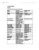
In the table this table below I have described the types of movement at each joint, showing the articulating bones, joint type and the agonist at each joint.
The Muscular System
- Muscles make up the bulk of the body and account for about one-third of its weight. Their ability to contract not only enables the body to move, but also provides the force that pushes substances, such as blood and food, through the body. Without the muscular system, none of the other organ systems would be able to function.
- The Skeleton and its joints support, protect, and provide flexibility for the body, but the skeleton cannot move itself. The job of that is performed by the MUSCULAR SYSTEM.
-
THE MUSCULAR SYSTEM is composed of muscle tissue or muscle fiber that is specialized to contract or shorten to produce movement during exercise or when needed.
Breakdown of a Skill
I will be breakdown down a skill to compare it from when a professional does the skill to when an amateur performs the skill, for this I will break down the football there are in stages to a strike the preparation, doing the strike and the follow through.
When learning the striking techniques there are a number of points to remember:
Strike Preparation
- Start by firstly selecting the target you want the ball to go in.
- Position yourself so the ball is directly between you and your target. You should be facing the ball and not turned sideways to it. Place your non-kicking foot (plant foot) along the side of the ball you feel more comfortable with, the toes of your plant foot can be toward the middle or back of the ball.
- Using slight flexion of the knee keep your non-kicking foot a little bent, so as to keep balance the muscles used will be the quadriceps, hamstrings and gastrocnemius
- Using flexion of the knee joint pull the kicking foot back creating a slight angle between you and the ball, using the muscles of the quadriceps and the hamstrings.
- Use lateral rotation at the hip to bring the kicking foot back to give more power behind the ball and making the body shape more open, the muscle used would be the latissimus dorsi and gluteus maximus.
- Flexion and extension at the arms will be present so as to keep balance, depending on what leg you are kicking with, the opposite arm will be flexed in front of the body using the biceps brachii and the other arm will be extended using the triceps brachii.
- Lastly the shoulder of the kicking foot will use lateral rotation to open up the body, so ready to kick the ball, the trapezius, pectorials major and deltoid are the muscles used in this motion.
The Pass
- The kicking foot should now be brought forwards show extension of the leg, muscles used would be the quadriceps, gastrocnemius; the kicking foot point downs in a plantarflexion position of the ankle the muscle used being the tibialis anterior
- The ball should be kicked with the laces part of the foot in the centre of the ball to make it accurate and get lots of power behind the ball, extension will still be present at the knee and plantarflexion at the ankle.
- Arms should switch so that the opposite arm to the kicking foot is in a extension position, so to give balance and kicking foots arm in flexion position using both the biceps brachii and triceps brachii in the arms.
- The kicking foots shoulder should now draw into the body using medial rotation, muscles used being the deltoid, trapezuis and the pectorials major.
- The hip should do exactly the same and draw in towards the body showing medial rotation using the quadriceps.
Follow Through
- Follow through with the kicking foot, so as to extend the knee and place all weight on to that leg to keep balance.
- Back leg should in a plantarflexion position using the muscle of the tibialis anterior
- Arms should remain in the same positions as before using the same muscles.
- Hip of kicking foot should be in mid-line of the body as performed by medial rotation.
Before During After
Muscle Groups
There are three muscle groups:
- Skeletal Muscle (Voluntary)
- Smooth Muscle (Involuntary)
- Cardiac Muscle (Cardio Vascular)
Each muscle type is different in the way they work:
Skeletal Muscle:
- These are activated by nerve stimuli to produce movement
- They comprise 40% of our body weight, and there is over 600 muscles
- They function in groups to accomplish sporting movement with great efficiency.
-
They are strained muscle
Cardiac Muscle:
- This is a highly specialized muscle only found in the heart
- It is the only muscle which is involuntary (works on its own)
- Made up of branched fibres giving it a striped appearance
- Contraction is controlled by a complex nervous and chemical system
- Resistant to fatigue
Smooth Muscle:
- These are involuntary muscles
- They are made up of spindle shaped cells
- Examples of these would be the muscles of the bowel, uterus and bladder
Muscle Contraction
Isometric Contraction—Muscle Actively Held at a Fixed Length
Isometric contraction is when the muscle is activated, but instead of lengthening or shortening, it is always held at a constant length. An example of this would be carrying an object in front of you. The weight of the object would be pulling down, but your hands and arms would be opposing the force with equal force going upwards. Since your arms are either raising or lowering, your biceps will be isometrically contracting.
The force generated during an isometric contraction is wholly dependant on the length of the muscle while contracting.
Concentric Contractions
When a muscle is working and required to lift a load which is less than the tension it can hold, the muscle begins to shorten. Contractions that permit the muscle to shorten are referred to as concentric contractions. An example of this would be raising the weight in a bicep curl.
Eccentric Contractions
When muscles are working they often are lengthening. Examples of this are walking, when the quadriceps (knee extensors) are working just after heel strike while the knee flexes, or setting an object down gently (the arm flexors must be working to control the fall of the object).
Eccentric contraction occurs when the load on the muscles increases, when it reaches a point where the external force on the muscle is greater than the force that the muscle can hold. This means that even though the muscle will be working fully, it is forced to lengthen due to the high pressure of the load.
Ligaments and Tendons
Ligaments
Ligaments are fibrous bands or sheets of connective tissue linking two or more bones, cartilages, or structures together. One or more ligaments provide stability to a joint during rest and movement. Excessive movements such as extension or flexion may be restricted by ligaments. Some ligaments prevent movement in certain directions.
Tendons
A tendon is a tough and flexible band of fibrous tissue. The tendon is the structure in your body that connects the muscle to the bones. The skeletal muscles in your body are responsible for moving your bones, thus enabling you to walk, jump, lift, and move in many ways. When a muscle contracts, it pulls on a bone to cause this movement. The structure that transmits the force of the muscle contraction to the bone is called a tendon.
Levers
First Order Levers
-
Fulcrum- the point of the movement or pivot, usually the centre of a joint in the body.
-
Load- the body weight or some external object
-
Effort- a muscular force to move the load
The fulcrum is between the effort and the load:
An example of this would be the load the neck has to carry the skull:
Second Order Levers
The load is between the fulcrum and the effort; the lever has a relatively small force and can move a larger weight. This is transmitted through the very strong achilles tendon.
Third Order Levers
The effort is between the load and the fulcrum. This is not as efficient as a second order lever, but a small muscle movement creates a long lever movement.
Agonist and Antagonist
When looking at the movement of the body it is important to consider the muscles. When movement takes place it is the prime movers (agonist) contract to allow movement to take place. The antagonists are the muscles that are resisting movement and so relax. So in the case of bending the arm at the elbow the prime mover (agonist) would be the biceps, and the triceps act as antagonists.
These types of muscles always in pairs that work against each other, and when one contracts the other relaxes.
Synergists
They assist the agonist and antagonist and fixator muscles, providing stability. The synergists reduce the amount of unnecessary movement when the prime mover contracts. They help to fine tune the movement.