In the early stages of osteoarthrisis, the chondrocytes proliferate forming clones. This is accompanied by biochemical changes as the water content of the matrix increases and the concentration of proteoglycans decreases. Subsequently vertical and horizontal fibrillation and cracking of the matrix occur as the superficial layers of the cartilage are degraded. Gross examination at the stage reveals a granular articular surface that is softer than normal. Eventually full-thickness portions of the cartilage are sloughed, and the exposed subchondral bone plate becomes the new articular surface. Friction smooth and burnishes the exposed bone, giving it the appearance of polished ivory. Concurrently there is thickening of the subchondrical bone plate and rebuttressing and sclerosis of the underlying cancellous bone. Small fractures through the articualr bone are common, and the dislodged pieces of cartilage and subchondral bone tumble into the joint forming loose bodies. The fracture gaps allow synovial fluid to be forced into the subchondral regions in a one-way, ball valve like mechanism. The fluid collection increases in size, forming fibrous walled cysts. Mushroom-shaped osteophytes develop at the margins of the articular surface and are capped by fibro cartilage and hyaline cartilage that gradually ossifies. The synovium shows minor alterations in comparison to the destruction of the articular surface and is congested and fibrotic and may have scattered chronic inflammatory cells.
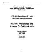
Pathological changes in osteoarthrisis.
(a) In a normal synovial joint the distance between the bone ends seen on radiography is maintained by the thickness of articular cartilage. (b) The early changes in osteoarthrisis is destruction of articular cartilage, which splits, become eroded and leads to narrowing of the joint space on radiography. There is inflammation and thickening of the joint capule and synovium. (c) With time, there is thickening of subarticular bone caused by constant friction of the two naked bone surfaces, leading to a highly polished bony articular surface. Small cysts develop in the bone beneath the abnormal articular surface causing irregular outgrowths of bone. ( From Stevens and Lowe Pathology 2nd edition)
Osteoarthrisis of the limb progresses slowly. Initially people feel discomfort in the limb joint. Usually this symptom would be felt for several years, which gradually gets worse with time, limiting the persons movement of the leg considerably. The onset is graduals with pain, limitations of movement and loss of function. The pain is greatest when the knee joint is used, or immediately after use, and is relieved by rest. Stiffness is less severe than in rheumatoid arthritis, and morning stiffness may not be as severe. As disorganization of the joint progresses crepitus may be felt or heard and joint effusions occur. As movement becomes more limited muscle wasting develops. Sometimes the lead into osteoarthrisis of the limb is due to osteoarthrisis in the hips. This is due to the reason that the pain fro the hip may often radiate to the knee joint. The limb joint is commonly involved as they become painful and tender. Flexion, valgus or varus deformities may occur leading to quadriceps wasting and marked effusion becoming present.
Treatment depends on the persons age and occupation as well as on the severity of the pain and stiffness. The natural history of the disease is extremely variable. Pain in osteoarthrotic limb joints may fluctuate in severity from time to time, and not infrequently subside to tolerable levels without treatment. Pain correlates poorly with the degree of stiffness and the severity of the disease as judged by radiographs, and the factors, which contribute to pain, are poorly understood. Limb joint stiffness is not always progressive and, unless it gets severe or several joints to get involved. It usually causes the person much less concern than the pain.
Radical surgical treatment is not often recommended and are advised not to be undertaken until conservative measures have been tried and the progress of the disease observed over a period of time. Conservative measures include reduction and avoidance of excessive stress on the affected limb joints. On the other hand, joint movement is necessary for the nutrition and health of articular cartilage and limitation of limb joint movement is one of the causes of cartilage degeneration, which may lead to or aggravate osteoarthrisis. Physiotherapy directed to the maintenance of joint movement by active exercise is of particular value in early osteoarthrisis of the limb and may prevent progression of the disease. Asprin may be prescribed to some patients to control pain in many cases. As symptoms vary from day to day, regular dose may be not necessary. Other anti-inflammatory and analgestic drugs may also be used to maintain pain however as they are pharmaceutical drugs patients are recommended to use them with doctors’ supervision. However no drug therapy yet known is had to have beneficial effect on the underlying disease.
In disabling osteoarthrisis of the limb possible surgical procedures may be of value. These are osteotomy, arthrodesis and replacement arthroplasty.
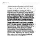
- In an osteotomy the bone is divided in the vicinity of a joint, and any joint deformity which may be present is corrected at the osteotomy site. The beneficial effect of such a procedure is easily understood which it corrects gross genuvarum or valgum, this restoring normal weight distribution within the knee. However, osteotomy of the upper end of the femur may relieve pain even when there had been no alterations in the alignment of the bone. Osteotomy may be indicated in early osteoarthrisis of the limbs in which pain is the predominant symptom and movement is not severely restricted.
Osteotomy of the tibia for osteoarthrisis of the knee
with varus deformity. Correction of faulty joint
alignment by osteotomy frequently relieves pain.
(From A companion to medical students in 3 volumes)
- Surgical arthrodesis of a joint gives complete relief of pain at the expense of movement. In the limb arthrodesis causes significant disability, but even in this site the operation may be indicated in a young patient, with advanced disease in one of these joints, whose occupation is physically heavy. Arthrodesis puts greater stress upon neighboring joints and should rarely be used when multiple joint involvements is present. Hence, two stiff knees would be a major handicap.
- Replacement arthroplasty is the substitution of an artificial joint for the diseased joint. This is a rapidly developing field of orthopedic surgery which hold great promise. Even the best of artificial joints, however, differ in one important respect from natural ones, in that they have no capacity for repair. For this reason wearing out or mechanical failure is a very real threat which dictates a certain caution in the use of replacement arthroplasty. So far it has been mostly used for management of chronic arthritis, particularly in the elderly and in patients with multiple limb joint disease.
In pure reality, there are still no satisfactory means of preventing osteoarthrisis in the limb nor are there any methods of halting its progression. This disease may stabilize for years at any stage but more often is slowly progressive over the remaining years of life. Although there are many attempts to find a possible cure for this increasing disease it has yet been found and so prevention is the best cure to osteoarthrisis of the limb today.
References:
Bolodeoku, J. Dogan, A. Holton, J.M. Lakhani, S.R. Lydyard, P.M. Patterson, K.G. Playfair, J.H. Pathology Integrated. Oxford Uni Press. London.2000
Collins, T. Cotran, R. Kumar, V. Robbins Pathologic Basis of Disease (6th edn). W.B. Saunders Company. Philadelphia.1999
Dirckx, J (ed). Stedmans Concise medical and Allied Health Dictionary. Williams and Wilkins. Baltimore USA. 1997
Garino, J. Lotke, P. Revision Total Knee Arthroplasty. Lippincott. New York. 1999
Passmore,R. Robson,J. A Companion To medical Studnets in 3 Volumes. Volume 3. Blackwell Scientific Publications. Oxford. 1984
Stevens, A. Lowe, J. Pathology (2nd edn). Mosby Publishing. London.2000
Stoller, D.W. MRI, Arthroscopy and Surgical Anatomy of the knee joints. Lippincott. New York. 1999