The axial skeleton main function is to protect and an example of this would be the cranium protecting the brain.
Some of the bones that are found in the axial skeleton are the cranium which consists of 8 bones and protects the brain and this is a flat bone.
The vertebrae column is also in the axial skeleton and this consists of 7 cervical vertebrae that are found in the neck, there are 12 thoracic vertebrae, which are in the chest area, and 5 lumbar vertebrae that are found in the lower back. These vertebrae protect the spinal cord that sends signals up and down your back and are irregular bones.
The ribs are part of the axial skeleton and there are 12 pairs in total these are flat bones, which are made to protect the organs within.
Some of the bones that are found in the appendicular skeleton are the scapulae’s that are flat bones and the clavicles that are short bones and these two bones make up your collarbones.
The arms are part of the appendicular skeleton and they are made up of the humerus, radius and ulna and all of these are long bones that are mainly used for movement.
Within the wrist there are 8 carpal bones which are short bones and in the hand there are 5 metacarpal bones and they are also short bones.
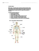
The phalanges are also found in the fingers and there are 14 in total, 2 in each of the thumbs and 3 in each finger these are shorts bones. (Figure 3 Appendicular)
The pelvic girdle and legs are found in the appendicular skeleton and contain an
illium, an ischium and a pubis and these are all there for support.
The femur, tibia and fibula are found in each leg and are there for support and movement, they come from the long bone category, also the patella is included into the leg and this is a sesamoid bone that is in the tendon of the knee.
The ankle and foot are made up of 7 tarsals and 5 metatarsals and they are short bones.
When playing and competing in sports your body will almost always be using movement.
To make movement happen the bones in your body act as levers, protection and support, all of these are what your bones have to do for the body to achieve the movement that it wants when playing sport.
When a goalkeeper in football uses an over arm throw he will be using his hands to grip the ball and this arm to put the force into launching the ball away from his body.
To do this he will use his humerus, radius, ulna, metatarsals and phalanges, the humerus then abducts and the radius and ulna extend, whilst this is happening the ball is being gripped by the hand and the phalanges are flexing around the ball.
When the ball is thrown the humerus adducts and the ulna and radius flex bringing the arm back into the body. At the last point of the movement the ball is released and the phalanges extend loosening the grip on the ball.
When kicking a ball in football you are using your foot and entire leg the bones used when kicking a ball are the tibia, fibula, femur, phalanges, tarsals and metatarsals. (Figure 4 Appendicular)
When you kick a football one leg is left for the support of the body whilst the other one is there to make the contact and put force onto the ball, if right footed the player would put all of the support onto his left leg whilst the right foot is off the ground.
The femur in the right leg would extend and then after that the tibia and fibula would follow also extending so that power is hitting the ball from the swing of the leg.
Then at the ankle plantar flexion occurs and the foot is brought into the football and the phalanges, tarsals and metatarsals are used to make contact with the ball.
You might also use your arms when kicking a ball as they can act to balance the body whilst on one leg, to do this the arms are normally out stretched and raised to shoulder length.
When this happens the humerus, ulna and radius are abducted outwards away from the body and this will make it easier to concentrate on kicking the ball and not balancing the body.
When in a game of football the ball made to be brought down to the ground with the help of the chest to do this the chest has to pushed outwards and the shoulders raised slightly whilst the arms can be brought out to help balance the body.
The bones that are used in this movement are the sternum, ribs, humerus, ulna, scapula and radius.
When the ball comes down to the body the first thing that it makes contact with is the ribs and sternum and there main function in this is mainly for protection of the vital organs behind them but it can also be used to stop the ball and bring it to the ground sp that it can be controlled in a quick movement. Whilst this happens the scapulas in the shoulders are elevated as the ball makes contact with the chest area and then quickly depressed to put a downwards force on the ball to bring it to the ground.
To help with the balance of the body the arms are used with the humerus flexing and the ulna and radius extending at the same time.
Task 2
a)
A joint is where two bones meet together, and some joints allow a large movement factor where as others are slightly movable, and some allow no movement.
Immovable joints also known as fibrous are all joined together by very strong fibrous connective tissue, and that is why they are not there for movement, a good example of this would be the cranium.
Slightly movable joints also known as cartilaginous joints have cartilage between the bones, which allow a small amount of movement and a good example of this would be the sternum and the clavical.
Movable joints are mostly known as synovial joints, and these allow great range of movement because they have synovial fluid which acts as a lubricant.
b)
Synovial joints will almost mainly be found in the appendicular skeleton as they are needed for greater range of movements in the arms and legs etc, and the cartilaginous joints will be mainly found in the axial skeleton as they are not needed as much for movement prepuces, and act more as protection and support.
There are six types of synovial joints, and they all have different (Figure 5 Synovial)
ranges of movement as some have large amounts of movement and others only allow small amounts.
Ball and Socket:
A ball and socket joint allows a huge range of movement, and can use such movements as flexion, extension, adduction, abduction, circumduction and rotation, and some examples of these would be found in the hip and shoulder.
A sporting example of this would be a footballer taking an over arm throw, the shoulder is circumducted to bring the ball back over the players head so that power can be forced into the ball.
Gliding:
At a gliding joint one bone slides over another, to create a small amount of movement, an example of this would be between the vertebrae and the carpals and tarsals.
Condyloid: (Figure 6 Ball and Socket)
This joint is very similar to a ball and socket joint but the only movements that can happen at it is flexion, extension, adduction, and abduction, and a good example of this would be at the wrist.
A sporting example of this joint would be a basket ball player taking a free throw and aiming to get a ball in the basket, to do this he must flex his wrist with the ball balancing in his hand and then push up causing extension at the wrist to get the back spin on the ball for it to hit the back board and go in.
Hinge: (Figure 7 Gliding)
At this joint the movement works just like a hinge, and allows movement back and forth (flexion and extension) an example of this would be the knee and the elbow.
A sporting example of this would be a ten- pin bowler bowling a ball, the arm is brought back into extension to gain the power to put into the ball to hit the skittles and then just before the ball is released the arm comes back into flexion to bring the ball to the ground.
Pivot:
With this joint it will only allow rotation, and can be found between the atlas and axis in the neck.
A sporting example of this would be a footballer putting spin onto a header to make the ball go into a certain place, and to do this he would have to make contact with the ball and his head and then quickly rotate his neck so that the head of the player just swipes the ball putting spin onto it.
(Figure 8 Hinge)
Bibliography
Figure 1 Bone Groups –
Figure 2 Axial Skeleton –
Figure 3 Appendicular Skeleton -
Figure 4 Appendicular Skeleton -
Figure 5 Synovial Joint –
Figure 6 Ball and Socket -
Figure 7 Gliding -
Figure 8 Hinge -