Pivot joints, these allow only rotation. A good example of this the neck, as a turn of the head can make a big difference in a football game. When a midfielder is considering his options they need to turn their head to look for the best player to pass to. This requires the sternocleidomastoid and the levator scapulae to turn the head. The forearms can also rotate. Turning the thumb outwards is called pronation, turning the thumb inwards is called supination.
Freely gliding; the metacarpal and carpals are a good example of this. Freely gliding joints only slide past each other. Carpals and metacarpals allow for a slight curve in the hand this can make a big difference when grabbing an object for example a football when the goal keeper hold the ball before a drop kick it would be hard to hold with out that slight curve in the hand. Dancers also need freely gliding joints a the tarso-metatarsal joints can also curve they require this to be able to support them self on tip toes for some dance moves.
Immovable Joints
Immovable joints are held together by actual intergrowth of bone or by strong fibrous cartilage. Immovable joints are rigid and they do not move hence the name. They are used in the skull to joint together parts of the skull and are fused in a way by intergrowth. These are fontanels. On the site that the sternocleidomastoid originates from the skull there is a fontanel called the mastoid fontanel. The Frontal bone, parietal bone, temporal bone and sphenoid bone are all fused joined by the sphenoid fontanel. These intergrowths are very important as in any sports it is easily possible to injure the skull especially in boxing with out these intergrowths the skull bones would easy move about and puncture the brain. For example a flurry of punches to the face in a professional boxing match would most likely kill a person due to the lack of intergrowths.
Slightly Movable Joints
These types of joints make up the front of the pelvis and the spine. They do not move much at all, and only move as if they did not this would cause the bones some physical stress. Slightly movable joints are held together by elastic cartilage. Slightly movable joints are also called cartilaginous joints. Each of the bones rests on a cushion of cartilage.
The bones can move slightly, but ligaments stop them moving too far.
An example of a slightly movable joint is the joint between two vertebrae.
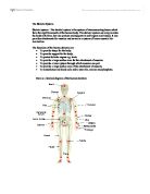
The Appendicular Skeleton
Anterior view of whole skeletal system
Taken from:
Above view of the Wrist and the Hand
Bones of the hand
2 examples of Carpals
Trapezium is an irregular bone as it is of an irregular shape. It can be felt in your hand by a deep groove. It is at the external and inferior part of the carpus it is in-between the meta-carpal bone and the radius. The superior surface is concaved and smooth. The inferior surface is ovaled in shape and concaved from side to side and convex from top to bottom. The anterior surface is narrow and rough. The upper part has a deep groove running from above downward and inward. The trapezium and the surrounding bones are all freely gliding (Dr. Gray, Henry, 1901 p 163). The grooves, concaves and convex shapes help other bones to glide past or move around it. Due to its irregular shape and its many neighbouring bones if this bone was to be injured during exercise for example if a boxer broke this bone as a result of a very hard counter-punch or other punch, it would be very painful to cope with and could take a considerable amount of time to heal because of it importance in day to day usage and exercise, if it is not properly rehabilitated and healed this can limit flexibility and strength of hand. It would also be open to further injuries and set backs in an athletes training programme and competitive events, especially if tissue was to heal into a fracture.
Articulates with, the scaphiod, trapezoid, second meta-carpal and the first meta-carpal.
Trapezoid is the smallest bone in its row. It has a Wedge shaped structure. It has a broader end forming a dorsal and a narrower end forming the pulmar. The superior surface is quadrilateral in from also smooth and slightly concaved it articulates with the scaphiod (another carpal). Is inferior surface articulates with the upper end of the seconds metacarpal, the trapezoid is convex from side to side. Its external surface is smooth and convex articulates with the trapezium. (Dr. Gray, 1901 p163).
This bone is also very important in day-to-day activities, exercise and sport. The concaved groove for the scaphiod is very important in gripping, as it moves during gripping. It is important for holding a tennis racket for example. It is a freely moving jointed bone.
Articulates with, the scaphiod, trapezium, second meta-carpal and the oz magnum.
Metacarpals There are five metacarpals in either hand, they are long bones and cylindrical in shape.
Common Characteristics of metacarpals; the shaft is a prism shape and it is curved at a longitudinal angle. It has 3 surfaces two of which are lateral and the other posterior. Both lateral surfaces and concave for the interossei muscle to attach and are separated by a prominent ridge the posterior surface is smooth, triangular flattened area, which is covered in tendons of the extensor muscles. Two lines beginning small tubercles on the dorsal either side bind it. Carpal bases are cuboid shapes and are broader at the front it articulates with the carpus and on each side with the neighbouring metacarpals; its dorsal and palmar surfaces are both rough so they can attach to tendons and ligaments. Carpal heads have a rectangular surface. They are convex and flattened laterally; it articulates with the proximal phalanx; it is also longer in some parts of it diameters than others. On either side of the head is a tubercle for the attachment of the lateral ligament of the metacarpo-phalangeal joint (joint of the metacarpal and phalange). The posterior surfaces are flattened and broad they support the extensor tendons; the anterior surface has a groove throughout in the middle for the flexor tendons to lay in. (Dr. Gray, 1901 p165)
The form and structure of the metacarpals and their joint types allow for your hand to appear to bend this help to grip an object, for example grip a tennis ball when serving with out the type of joint and the long cylindrical structure of the metacarpals it would be impossible for our hand to grip.
Picture of hand Taken from
Phalanges of the hand they are the tips of the fingers and are split into three rows the first row being the proximal Phalanges are vital in a lot of sports as they can wrap around a ball or object involved in sports and exercise. For example when a tennis player serves the ball the ball is secured in the hand with the fingers metacarpals. They are adapt for good gripping properties due to their shape making them good for securing the tennis ball the metacarpals in the hand only allow for flexion and extension because their joints are hinge joints this movement being restricted to only flexion and extension allows for a controllable grip for tennis as the fingers will not abduct and adduct.
Anterior View of the Arm and Shoulder
Taken from
The Clavicle, it can be felt throughout its entire span in any human regardless of obesity. At the inner end is the enlarged sternal extremity where the bone is above the upper margin of the sternum. This can also be felt quite easily forming with the sterno-mastiod a V-shaped notch, the pre-sternal notch. Passing out ward is the shaft of the bone that can be felt immediately under the skin. The clavicle or collarbone is at the anterior part of the shoulder. The clavicle is a long bone that has a curve in its shaft. It can be found placed horizontally at the anterior view of the thorax just above your first rib. Its articulations are with the inner extremity of the sternum and by its outer extremity with the scapula, allowing for great latitudinal motion in the arm.
(Dr. Gray, 1901 p135-137). The clavicle helps to protects the humerus.
The flattened portion; the outer third is flattened from above and downwards giving it two surfaces; upper & lower also two borders; anterior & posterior. The upper surface is flat and roughed it is marked by impressions of the deltoid attachment in front and impressions are also made by the trapezius behind. In-between those two impressions is a small part of bone, the under surface is flattened. At its posterior border is the conoid tubercle it surmounts the coracoid process of the scapula and gives attachment to the conoid ligament. The anterior border is concave and thin also rough it gives attachment to the deltoid. It presents at its inner end a tubercle called the deltoid tubercle. (Dr. Gray, 1901 p135-137)
The internal portion; part of the clavicle that is prismatic in forms the inner two thirds of the clavicle it is convex in front meaning it is concave behind, marked by three borders, therefore presenting three surfaces, the most anterior border uninterrupted with the anterior margin of the flat portion. At its start the surface is smooth corresponding to the interval between the attachment of the pectoralis major and deltoid muscles. The inner part of the clavicle forms an elliptical part for attachment. (Dr. Gray, 1901 p135-137)
Anterior View of the Pelvis and Leg
Taken from
Femur: The shaft is almost a perfect cylinder. A little broader at the bottom than at the top. At the top part of the femur the ‘head’ there is a Synovial joint; a ball and socket joint allowing articulation with the pelvis. At this joint all types of movement are possible; rotation abduction addiction flexion and extension. The surface of the femoral head is smooth apart from a small groove called the fovea capitis femoris This joint is held together with tendons to connect the bones and tendons around the skeletal muscles. Because of its wide range of movement it should be easily dislocated for example receiving a bad tackle to the inside thigh of your leg in theory should dislocated the femur, but because of the surrounding muscles almost constant daily use the surround muscles, tendons and ligaments are very strong making it difficult to dislocate. The femur is the longest and strongest bone is the human body. The body of the femur presents three borders: posterior that corresponds to the linea aspera, lateral and medial. These borders delimite three surfaces: anterior, posteromedial and posterolateral. The femurs shaft is slightly arched in that it is convex at the anterior view and concave behind; where some of its strength comes from a prominent longitudinal ridge called the linea aspera. The at the form a . The on the distal end, are bumps that fit into corresponding articular facets on the . The gap between the two condyles is called the (or notch). Above the femoral condyles are the medial and lateral ; above the medial epicondyle is the . ()
Patella: also known as the knee bone provides a blockade to obstruct the tibia and fibula (lower leg) from hyper-extending as the knee joint is a hinge joint it will only allow for flexion and extension, but the knee will only extend to a certain degree where the femur is roughly in line with the tibia. This bone is very useful especially for running athletes, as with out the patella this joint would be too ‘flimsy’ making it very difficult for you to support yourself in an intense sprint. The patella is triangular, it is known as a sesamoid bone. The patella also protects the joint behind and increases the leverage of the quadriceps extensor by giving it a greater angle to pull on. Its anterior surface is convex. The muscle is attached to the base of patella. The vastus lateralis and are attached to lateral and medial borders of patella. The patella’s leverage advantages can help for example footballers to kick a ball a lot harder.
Tibia the shaft of the tibia is a traingular prism in shape and broader at the superior end than the inferior end at the head of the fibula it enters the knee joint and is connected to the quadriceps via a ligament stretching over the patella. The lower and thinner part of the extremity is where fractures in sport occur most frequently. Its triangular primed shape allow for the bone to be more resistant to fractures and clean breaks which is very use full just a cylindrical shape in such a load bearing area would be prone to injury in sports for example its common for footballers to get a good kick in the shin the triangular form spread the stress making it more likely to bruise than fracture. The triangular form can also allow for deeper muscles it grow and increase in strength under the superficial muscles again allowing for resistance to injury and can also allow for better performance. Its lower extremity much small than the upper.
Fibula the fibula or calf bone is a placed on the lateral side of the , with which it is connected above and below. It is the smaller of the two bones, and, in proportion to its length, the most slender of all the long bones. Its upper extremity is small, placed toward the back of the head of the tibia, below the level of the , and excluded from the formation of this joint. Its lower extremity inclines a little forward, so as to be on a plane anterior to that of the upper end; it projects below the tibia, and forms the lateral part of the ankle-joint. The fibula also makes part of the knee joints and is necessary for proper stable movement in the legs is position helps to sufficiently gain a proportionate joint size in both the ankle and knee joints. Which is vital in a lot sports to keep balance for example runners need a good amount of balance to be able to keeps running as fast as they can as their C.O.M. shifts continuously the fibula role in knee and ankle joints its articulations and ligament and tendon connections help to keep balance. (http://en.wikipedia.org/wiki/Fibula)
Above View of the Foot
Taken from
Metatarsals: There are five metatarsals in either foot, they are long bones and cylindrical in shape. Like most bones are subdivided into a shaft and two extremities the ‘heads’ of the bone.
The shaft is a prism and tapers gradually from the tarsal above to the extremity closer the phalanges; they are also curved longitudinally making them slightly convex above and concave below. The posterior extremity is wedge shaped and articulates with the tarsals. The anterior extremity’s sides are flattened and present a depression surmounted by a tubercle.
The Meta tarsals basically serve for mobility in the foot and balance; they joint the toes to the foot (tarsal to the phalanges). Injury to the Meta tarsal can set back an athletes training programme and competitive involvement for some time. For example when Wayne Rooney broke his fifth meta-tarsal he missed several months of training and the first few games of the 2006 Fifa world Cup.
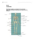
Axial Skeleton
Side view of the Spinal column
Taken from
Below picture scanned from Grays Anatomy (Dr. Gray, 1901 p150)
The spine; in total is made up of 33 irregular bones 9 of which are fused. The average length is about two feet measures along the curve. The spines many bones allow for the bending of the trunk controlled by the deeper trunk muscles. It is the spine that holds the torso upright and gives main support to the body. The spine is very important and strength of the spine bones and surrounding muscles. Its structure allows for the back to curve, which is important in such sports that require good trunk flexibility such as gymnastics for example trampoline requires the structure of the spine to bend the back on certain twists and turns. The structure of all the freely gliding joints in-between the spinal columns bones. The Spinous process helps for leverage for the muscles and tendons to connect to the vertebrae. Allowing for a more energy efficient contraction.
Taken from ()
The Skull
Taken from
The upper skull is primarily for the purpose of protecting the brain as the slightest amount of trauma can render an athlete concussed or unconscious. It is rounded to accommodate the brain. And articulates with the first vertebrae (C1) of the cervical vertebrae section of the spine and the mandible (jaw) strength of the skull Is important in almost every combat sport e.g. boxing as in boxing a lot of the punches landed are to the head in an attempt to knock the opponent dizzy, the mandible also needs considerable strength as the ‘knockout punch’ is a punch in the chin which can force the head back and cut of oxygen to the brain. A weak mandible would break easily after receiving a knock out punch from a professional boxer.
On the exterior the cranial bones include the two frontal bones, which make up the forehead and which fuse together in adulthood; the two parietal bones, which make up the top of the head and which in early childhood are separated from the frontal bones by a space called the anterior fontanelle; the single occipital bone, constituting the back of the skull, which is pierced by a large opening through which the spinal cord enters the cranial cavity; and the two temporal bones, which make up the temples and side of the head and bear the zygomatic processes, or cheekbones.
Internally, the cranial bones include the ethmoid bone, which forms part of the septum of the nose and through which the olfactory nerves pass from the brain to the upper and middle turbinates; the sphenoid bone, which constitutes most of the floor of the cranial cavity and which houses the pituitary gland; and part of the occipital bone. The floor of the cranial cavity contains three terraced depressions, which contain the cerebellum and the frontal and temporal lobes of the cerebrum.
The facial bones include the two nasal bones, which constitute the upper portion of the bridge of the nose; the two lacrimal bones, which are located in each eye orbit next to the nose, close to the tear ducts; the maxillary bone, which constitutes the upper jaw; the mandible, which constitutes the lower jaw; the two palatine bones of the hard palate; the vomer, which, with a part of the ethmoid bone, constitutes the nasal septum; and the two inferior turbinates of the nose.
Anterior View of the Pelvis
Taken From
The pelvis is roughly symmetrical and each side is actually made up of three separate bones:
-
the upper half (the broad "wings") is the
-
the middle (the top half of the lower "loops") is the
-
the bottom (the lower half of the "loops") is the
These three bones fuse together with age and are collectively known as the hip bone, so coaxes, or the in nominate bone. The pelvis is joined to the bone by (the ), and the bones nest in specially shaped sockets (the ) on each side. The upper edge of the ileum is known as the iliac crest. The place at the front of the pelvis where both pubis bones join together is called the . This is normally a very inflexible joint, but it softens and becomes more flexible during late pregnancy, allowing it to expand during labour for the baby's head to pass through.
()
Pelvis, lower part of the trunk of the body, bounded at the front and on either side by the hipbone and at the back by the sacrum and the coccyx, the lowest part of the spinal column. The hipbone is composed of three separate bones: the ilium, the ischium or lower part of the hipbone, and the pubis, the central pubic bone that unites with the ischium at either side. In early life, the three bones are separate, then in late teens or early 20s they unite to form a single structure called the innominate bone. At the lower end of the hipbone is a cup-shaped depression called the acetabulum in which the femur, or thighbone, rotates. The pelvis thus acts as a unit in all bodily movements. The weight of the trunk is transferred from the spine through the sacrum and then through the hipbone to the thighbone and the lower extremities. Conversely, all forces acting on the lower limbs are transmitted to the trunk by the same route.
Sacroiliac Joint, fibrous joint in the lower back between the lumbar vertebrae and the coccyx, the bones at the lower end of the spinal column. It is composed of five fused vertebrae that form a solid triangular bone (the sacrum), and the ilium parts of the two hipbones. Once believed to be the site of sprain and lower back pain, powerful ligaments that avert injury in fact bind it. A displaced vertebral disc more generally causes such pain. Taken from: Microsoft ® Encarta ® Encyclopaedia 2005 © 1993-2004 Microsoft Corporation. All rights reserved.
The structure of the pelvis allows for the hips a (ball and socket joint) to be present to allow for all types of movement which is useful in many sports especially in rhythmic gymnastics as practically every movement possible is required, e.g. the splits require you to abduct both legs then adduct a front kick required flexion of the hips and a spin may require rotation, all of which supplied by the ball and socket joint.
This wide range of movement has applications to many sports and exercise examples. The ball and socket joint in the hip is supported in the pelvis, this allowing for all types of movement meaning that a person can side step walk backwards forwards and rotate their leg. In football all of these types of movement are needed, the pelvis and femur ball and socket joint when running to tackle a player, needs to flex and extend and if the player needs to quickly move out of the way of someone else they need to abduct then adduct their legs one after the other to be able to side step out of the way.
Reference List:
Text references;
Greys anatomy, 1901, Dr Henry Gray
Microsoft ® Encarta ® Encyclopaedia 2005 ©
Image references;
Greys anatomy, 1901, Dr Henry Gray
Christopher Carter, Unit Seven Anatomy For Sports and Exercise Science, Skeletal System and joints