The ribs (flat bone) -there is 26 ribs in total, 2 of which are floating ribs and these are used in the skeleton system to help protect the vital organs such as the heart and the lungs etc… It has its shape because it needs to be hollow to house the organs and it also allows movement for when the lungs inhale and exhale air.
The bones in the appendicular skeleton are:
Clavicle (flat bone) -In human anatomy, the clavicle or collar bone is a bone that makes up part of the shoulder girdle (pectoral girdle). It receives its name from the Latin clavicula (little key) because the bone rotates along its axis like a key when the shoulder is abducted
The clavicle serves several functions:
It serves as a rigid support from which the scapula and free limb are suspended. This arrangement keeps the upper limb away from the thorax so that the arm has maximum range of movement but at the same time makes sure that the humerus does not dislocate as easily as it potentially could. It also Transmits impacts from the upper limb to the axial skeleton.
Even though it is classified as a long bone, the clavicle has no medullary (bone marrow) cavity like other long bones. It is made up of spongy (cancellous) bone with a shell of compact bone. The clavicle bone is the only bone that does not have marrow. It is a dermal bone derived from elements originally attached to the skull.
Scapula (long bone)-In anatomy, the scapula, or shoulder blade, is the bone that connects the humerus (arm bone) with the clavicle (collar bone).
The scapula forms the posterior part of the shoulder girdle. In humans, it is a flat bone, roughly triangular in shape.
Humerus (long Bone)-The humerus is a long bone in the arm or fore-legs (animals) that runs from the shoulder to the elbow. On a skeleton, it fits between the scapula and the radius and ulna. It consists of the following three sections:
Upper extremity of humerus
Body of humerus
Lower extremity of humerus
This is used to support the upper arm and is the catalyst for movement in this part of the body.
Radius (long bone) -The radius is the bone of the forearm that extends from the inside of the elbow to the thumb side of the wrist. The radius is situated on the lateral side of the ulna, which exceeds it in length and size. Its upper end is small, and forms only a small part of the elbow-joint; but its lower end is large, and forms the chief part of the wrist-joint. It is a long bone, prismatic in form and slightly curved longitudinally.
Ulna (long bone) -The ulna (Elbow Bone) is a long bone, prismatic in form, placed at the medial side of the forearm, parallel with the radius.
Carpals (sesamoid bones) -The carpals consists of seven bones arranged into two rows. There are three bones in the proximal row. The radial carpal is on the medial side and is joined proximally to the radius. It is the largest of the carpal bones.
Metacarpals (sesamoid bones)-There are five metacarpal bones. They are long, slender bones with relatively large extremities. The proximal extremity is called the base and the distal one is the head. They are numbered from medial to lateral like the carpal bones. Each articulates with its corresponding carpal bone except the fourth and fifth which both articulate with the fourth carpal bone. Distally they all articulate with the corresponding proximal phalanges.
Phalanges (short bones) - The name Phalanges is commonly given to the bones that form fingers and toes. In primates such as humans and monkeys, the thumb and big toe have two phalanges, while the other fingers and toes consist of three. Phalanges are classified as long bones.
The phalanges do not really have individual names but are named after the digit, and their distance from the body.
Distal phalanges are at the tips of the fingers and toes.
Proximal phalanges are closest to the hand (or foot) and articulate with the metacarpals of the hand, or metatarsals of the foot.
Middle or Intermediate phalanges are between the distal and proximal. The thumb and big toe do not have middle phalanges.
The term phalanx or phalanges refers to a Greek army formation in which soldiers stand side by side, several rows deep, like an arrangement of fingers or toes.
Pelvic gurdle (flat bone)-The pelvis (pl. pelvises or pelves) is the bony structure located at the base of the spine. The pelvis incorporates the socket portion of the hip joint for each leg.
The pelvis is symmetrical and each side is actually made up of three separate bones:
the upper half (the broad "wings") is the ilium
the middle (the top half of the lower "loops") is the pubis
the bottom (the lower half of the "loops") is the ischium
These three bones fuse together with age and are collectively known as the hip bone, os coxae, or the innominate bone. The pelvis is joined to the sacrum bone by ligaments (the sacroiliac joint), and the hip bones nest in specially shaped sockets (the acetabulum) on each side. The upper edge of the ilium is known as the iliac crest. The place at the front of the pelvis where both pubis bones join together is called the symphisis pubica. This is normally a very inflexible joint, but it softens and becomes more flexible during late pregnancy, allowing it to expand during labour for the baby's head to pass through.
Sexual differences
A female pelvis is also wider and shallower than a male pelvis. A well-known way of determining the sex of a pelvis is to compare the angle of the width of the frontal opening to one's hand.
If the angle is about the same as between the outstretched thumb and index finger, it is a female pelvis (arcus pubis).
If it is closer to the angle between the spread index and middle fingers, it is a male pelvis (arcus subpubis).
Function
The pelvis protects the digestive and reproductive organs in the lower part of the body, and many large nerves and blood vessels pass through it to supply the legs. It is also an important load-bearing part of the skeletal system.
Femur (long bone) -The femur or thigh bone is the longest, most dense, and strongest bone of the human body. It forms part of the hip and part of the knee.
The word "femur" is Latin for "thigh".
The femur consists of a head and a neck proximally, a (or shaft), and two ( and ) distally
Proximal end
The femur's head forms a ball-and-socket joint at the hip.
Other proximal features of the bone include the greater trochanter and the lesser trochanter, two bony projections that allow muscles to attach.
Posteriorly the gluteal tuberosity is a rough surface that gluteus maximus attaches to. Beneath this, the linea aspera runs down the back of the femur, which also provides an attachment for the biceps femoris muscle.
One important function of the femoral head is the production of red blood cells within the bone marrow.
Distal end
Distal end of the femur
The condyles at the knee form a condylar joint.
The medial and lateral condyles on the distal end, are bumps that fit into corresponding articular facets on the tibia. The gap between the two condyles is called the intercondylar fossa (or notch). Above the femoral condyles are the medial and lateral epicondyles, above the medial epicondyle is the adductor tubercle.
Tibia (long bone) -The tibia is the larger of the two bones in the leg below the knee in humans and other vertebrates.
The tibia or shin bone, in human anatomy, is found medial (towards the middle) and anterior (towards the front) to the other such bone, the fibula. It is the second-longest bone in the human body, the largest being the femur. The tibia articulates with the femur and patella superiorly, the fibula laterally and with the ankle inferiorly.
Gender differences
In the male, its direction is vertical, and parallel with the bone of the opposite side, but in the female it has a slightly oblique direction downward and lateralward, to compensate for the greater obliquity of the femur.
Fibula (long bone) -The fibula or calf bone is a bone placed on the lateral side of the tibia, with which it is connected above and below. It is the smaller of the two bones, and, in proportion to its length, the most slender of all the long bones. Its upper extremity is small, placed toward the back of the head of the tibia, below the level of the knee-joint, and excluded from the formation of this joint. Its lower extremity inclines a little forward, so as to be on a plane anterior to that of the upper end; it projects below the tibia, and forms the lateral part of the ankle-joint. Its function is to support the leg and also to help with muscle attachment in the leg/calf
Patella (sesamoid bone)-The patella or kneecap is a thick, triangular bone which articulates with the femur and covers and protects the front of the knee joint. It is the largest sesamoid bone in the human body. It is attached to the tendon of the quadriceps femoris muscle, which contracts to straighten the leg..
The primary functional role of the patella is knee extension. The patella increases the leverage that the tendon can exert on the femur by increasing the angle at which it acts.
The patella ossifies (starts to solidify) between the ages 2-6 years. In some people it may be absent congenitally or hypoplastic. In 2% of the population there is a bipartite patella, which is usually asymptomatic.
Tarsals (short bone)-In humans, the tarsals are the cluster of bones in the foot between the distal ends of the tibia and fibula and the metatarsals. The bones of the tarsals do not belong to individual toes, whereas those of the metarsals do. The joint between the tibia and fibula and the tarsus is called the ankle. this bones shape is adapted to it function via its structure because it is quite a small group of bones if u just take one and analyze it then you will find that it would be a hard bone to break due to a force being applied above or from below as it is covered in compact bone. It is also in a good shape to help with fluid movement.
Metatarsals (short bone)-The metatarsals consists of the five long bones of the foot, which are numbered from the medial side, each presents for examination a body and two extremities. Metacarples are the part which forms the long part of the foot.this bones shape is adapted to it function via its structure because it is quite a small group of bones if u just take one and analyze it then you will find that it would be a hard bone to break due to a force being applied above or from below as it is covered in compact bone. It is also in a good shape to help with fluid movement.
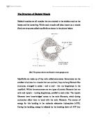
The Structure of a bone.
Bones are one of the only organisms in the body that can repair itself if it gets damaged as it is constantly damaging and renewing itslef . This renovation of the bone occurs at a accelerated scale when the bone is growing. The bones holds certain salts and minerals. These are calcium carbonate and calcium phosphate. It is these that give bone its well known properties. Also collegen fibres give bones the ablity to bend making it less brittle.
There are a lot of bones in human anatomy but they are all made up of two types of essential bone tissue. These are:
Compact or cortical tissue
This type of tissue is the so called armour of the bone. The way compact bones is structured it is built to bear the weight of the body and protect it from breakages through the long bones.
Cancellous or spongy bone
This is held in the epiphysis of a bone and this is where red blood cells are produced. This type of bone tissue looks like a sponge but the only thing is that it is not soft it is very hard.
All bones are inside a important membrane called the periosteum. This contains blood vessels to deliver the necessary foods for the bone to develop. Inside the bone and the periosteum there are 3 different types of cells and they are:
Osteoblasts
This is also known as the building blocks for bones. It produces collagen and minerals (i.e. calcium) to assist with healthy development.
Osteocytes
This is the prinicpal cell for bone tissue and is improtant for healthy development of bones.
Osteoclasts
This type of bone can be reffered as the pac men of the bone and they remove parts of the bone they no longer see healthy or fit.
For everyone, they are not born with fully formed bones anywhere in the bondy they all start as cartilage. During infancy, calcium and phosphrous are laid and the bones start to eventually calcify (harden). A bone develops from the central parts out edge and has a honeycombed innner networked mesh and an outer compact layer of bone. Ossification is where the bone development occurs.