Smooth Muscle
Smooth muscle is non-striated and involuntarily controlled this means it moves without us needing to think about it. It is found in blood vessels and many of the digestive system organs.
Smooth muscle is made of single, spindle-shaped cells. It gets its name because no striations are visible in them. Each smooth muscle cell contains thick (myosin) and thin (actin) filaments that slide against each other to produce contraction of the cell. The thick and thin filaments are anchored near the plasma membrane (with the help of intermediate filaments).
Smooth muscle (like cardiac muscle) does not depend on motor neurones to be stimulated. However, motor neurones (of the autonomic system) reach smooth muscle and can stimulate it - or relax it - depending on the neurotransmitter they release (e.g. noradrenaline or nitric oxide, NO).
Smooth muscle can also be made to contract:
- by other substances released in the vicinity (paracrine stimulation)
Example: release of histamine causes contraction of the smooth muscle lining our air passages (triggering an attack of asthma)
- by hormones circulating in the blood
Example: oxytocin reaching the uterus stimulates it to contract to begin childbirth.
The contraction of smooth muscle tends to be slower than that of striated muscle. It also is often sustained for long periods. This is called tonus but the mechanism is not like that in skeletal muscle.
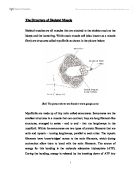
A magnified picture of smooth muscle is as follows:
(www.google.com)
Cardiac Muscle
Cardiac or heart muscle is like skeletal muscle in some ways: it is striated and each cell contains sarcomeres with sliding filaments of actin and myosin.
A picture of Cardiac Muscle is as follows:
(www.google.com)
Cardiac muscle also has a number of unique features that reflect its function of pumping blood.
- The myofibrils of each cell (and cardiac muscle is made of single cells and a single nucleus) are branched.
- The branches interlock with those of adjacent fibres by adherens junctions. These strong junctions enable the heart to contract forcefully without ripping the fibres apart.
- A heartbeat is generated within the heart itself. Motor nerves (of the autonomic nervous system) run to the heart, but their effect is simply to modulate - increase or decrease - the intrinsic rate and the strength of the heartbeat.
- The action potential that drives contraction of the heart passes from fibre to fibre through gap junctions.
- All the fibres contract in a synchronous wave that sweeps from the atria down through the ventricles and pumps blood out of the heart. Anything that interferes with this wave may cause the fibres of the heart to beat at random - called fibrillation.
- Cardiac muscle has little glycogen and gets little benefit from glycolysis so most of the energy needed is taken from oxygenated blood.
Skeletal Muscle
The main function of the skeletal muscle is to produce movement which is where it gets its name from (the muscle moves the skeleton which in turn moves our body)
A picture of skeletal muscle is as follows:
(www.google.com)
Motor Neurone
The Motor Neurone forms synaptic junctions with either extrafusal muscle fibres (skeletal muscle) or intrafusal muscle fibres (thread-like muscle that adjusts tension). Stimulation of these motor neurones induces contraction or shortening of the muscle fibres. Alpha motor neurones induce the contraction of extrafusal muscle fibres upon stimulation, whereas gamma motor neurones induce the contraction of intrafusal muscle fibres upon stimulation. Alpha motor neurones control muscle contraction involved in voluntary movement, whereas gamma motor neurones control muscle contraction in response to external forces acting on the muscle. In response to these external forces, the gamma motor neurones induce the involuntary, reflexive movement called the stretch reflex. Intrafusal motor neurones adjust the length of intrafusal muscle fibres to maintain an appropriate level of tension on the muscle spindle receptor. The control of intrafusal muscle fibres occurs independently of the length of skeletal muscle fibres. This independent function allows the spindle to maintain a high degree of sensitivity over a wide range of muscle lengths, and in effect acts as a means of encoding muscle length.
The smallest functional component of the motor system is the motor unit, which is composed of the motor neurone and all muscle fibres that it innovates (Parent, 1996). The innovation ratio defines the number of muscle fibres innovated by a single motor neurone. Low innovation ratios (approximately 100) occur in the hand, where fine gradations in muscle force are needed for fine motor control. Conversely, high innovation ratios (approximately 2000) occur in larger muscles such as the biceps of the upper arm where accuracy of movement is relatively insignificant.
When a motor neurone fires, the neurotransmitter acetylcholine is released in the neuromuscular junction. Depolarisation is propagated in both directions along the muscle fibres innovated until the ends are reached. Upon completion of this signal propagation, the muscle undergoes a brief contraction. The activation of motor neurones is modulated by local circuits in the spinal cord and by pathways descending from motor centres of the brain.
There are two types of gamma motor neurones that work in association with the two types of muscle spindle sensory fibres, the dynamic gamma motor neurone (involved in responses to dynamic stretch) and the static gamma motor neurone (involved in the response to steady-state length).
Reference Parent, A. (1996). Carpenter's human neuroanatomy (9th ed.). London: William’s & Wilkins.
Neuromuscular Physiology (What Sparks A Muscles Contraction?)
Muscle fibres are stimulated by the nervous system (motor neurones). Each muscle fibre is innovated by only one neurone. A neurone and the fibres it innovates are referred to as a motor unit. All of the muscle fibres in a motor unit are usually of the same fibre type. All of the fibres in a motor unit must fire or none of them.
How does the neurone 'innovate' its associated muscle fibres?
The neurone 'connects' to the fibres at their centre (their length-wise centre). To innovate them they transmit an electric current to the fibres, which travels out from the centre of the fibres to their ends, this sets off a contraction.
Muscle Firing Patterns
Muscle Fibres have two recruitment patterns. In the first pattern, units that innovate the same types of fibres are recruited at different times, so that some units are resting (recovering) while others are firing. Obviously, at high loads this pattern isn't possible because all available motor units will have to be fired at the same time to lift the load. In the second pattern, motor units that are more fatigue resistant are recruited before fibres that are more rapidly fatigued. (Taken from )
Muscle Fibre Types
Striated skeletal muscle comes in three different fibre types.
They are:
- Type I: slow twitch (ST), slow oxidative (also called red fibres)
- Type IIA: fast twitch (FT), fast oxidative (also called white fibres)
- Type IIB: fast-glycolytic (a kind of white FT fibres)
FT fibres have more access to myosin ATPase than ST fibres. This allows them to release energy more quickly and deliver more power than ST fibres. FT fibres are also larger in diameter because of higher concentrations of actin and myosin filaments within them as compared to ST fibres. This further allows them to develop more force. “ST fibres have greater intramuscular triglyceride stores (for sustained energy), more aerobic enzyme activity, more of a substance called myoglobin (which is instrumental in the process of using oxygen to create energy), greater mitochondrial density (mitochondria manufacture about 95% of the ATP that exists in muscle tissue) and greater capillary density”. ()
For the above reasons:
- FT fibres are best suited to generating large amounts of force over a short period and are sensitive to fatigue and ST fibres are best suited to low-load, long duration activities.
FT fibres, type IIAs has both good anaerobic and aerobic qualities. They have high ATPase activity like fast-glycolytic (IIB) fibres, but also a high oxidative capacity like type I fibres. Because of this, they can maintain a contraction longer than type IIBs, but contract faster (therefor developing more power) than type Is. Type IIBs do not exhibit this duality and are poor performers aerobically but very well equipped for anaerobic activities. They can, consequently, develop even more short-burst power than the IIAs. Both types of FT fibres have significantly larger innovating neurones than STs and, therefore, have higher activation thresholds than STs. They are activated only after the STs have been fired, but they can twitch faster and more often. FT fibres are brought into play by either the effort to more a heavy load or by the need to move an object faster than is possible with ST fibres. Type IIB fibres can twitch three times faster (and therefore, more often) than ST fibres. Type IIAs can also twitch faster and more often than ST fibres. Because of this, and the recruitment pattern, a FT fibre may begin its contraction after a ST fibre but actually finish at the same time or before. This leads to another contributor to the FT fibres abilities to produce greater force - their enhanced frequency of firing. Because they complete the firing sequence more quickly they can fire more often than ST fibres, thus developing more tension.
The force developed by a muscle depend on the number of fibres that are forced to contract (the more units contracting, the more force developed). A sudden increase in force is met by the involvement of more motor units. So a weight lifter will be using more motor units than a cross-country runner at any one time. If the weight lifter increased his weights to a degree that the units are under fatigue then they will increase the frequency of firing therefor meeting the muscles demand.