The nervous system was considered as research on birds, reptiles, amphibians and fish by Cassone (1990) was performed. The pineal gland, which regulates circadian rhythms through its release of melatonin, was then implicated. Although in most mammals, including humans, it does not have an effect on reproductive activity, it does in these due to the effect of light and dark in seasonal changes.
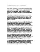
Figure 1: Control of the anterior and posterior pituitary by the hypothalamus
This, in turn, lead to the hypothalamus being an obvious option, as the pituitary was attached to this it made sense. Various experiments soon confirmed this, though how it was able to do was still not apparent, as there was no neural connection between the hypothalamus and the anterior pituitary, only the posterior pituitary, shown in figure 1.
The hormones vasopressin and oxytocin are released from the posterior pituitary after being synthesised in the paraventricular and supraoptic nuclei of the hypothalamus. Oxytocin would control contractions during labour and suckling of young, whereas vasopressin would aid water reabsorbtion in the kidneys. This could be explained easily, as there was a neural connection between the hypothalamus and the posterior pituitary.
The anterior pituitary, on the other hand, had no neural connection with the hypothalamus. Harris (1955) still hypothesised that this was regulated by the hypothalamus. The hypothalamopituitary portal system was soon discovered whereby capillaries in the hypothalamus transported information through portal veins to capillaries in the anterior pituitary. This was proven when cutting the portal vein meant hormone release was affected.
Once this connection was identified, it was suggested that there were specific chemicals in the hypothalamus that triggered the release or stopped the release of hormones from the anterior pituitary, called releasing hormones or inhibiting hormones respectively. Schally, Kastin and Arimura (1971) identified that gonadotropin-releasing hormone in the hypothalamus caused the release of gonadotropins from the anterior pituitary, namely follicle-stimulating hormone and luteinising hormone.
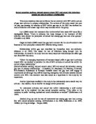
Figure 2: A summary model of the regulation of gonadal hormones
The hypothalamus therefore has much control over sexual behaviour. It governs the actions of the pituitary gland and, in turn, spurring the release of other hormones from the gonads, illustrated in figure 2. However, there are slight differences between the hypothalamus of the male and the female, defined above, which result in slight differences in its function.
An explanation for this difference was put forward by Gorski et al (1978) that there was an area in the medial preoptic area of the hypothalamus that was greater in size in males than in females, this was termed the ‘sexually dimorphic nucleus’ (Pinel, J. P. J. 2003. P.344). The rats are born with the same size nucleus, but the male sexually dimorphic nucleus then increases in size at a much more rapid rate then the female. The size of male rat’s nucleus is due to the levels of testosterone, as further tests by Gorski (1980), where male rats have been castrated and female rats injected with testosterone, have indicated.
There have also been a number of other differences between the hypothalamus of males and females discovered since. With respect to humans, there have been nuclei found in the preoptic, suprachiasmatic and anterior parts that are significantly smaller in females than in males. Therefore, when looking at the role the hypothalamus plays in sexual behaviour, we will consider males and females separately.
In males, the medial preoptic area of the hypothalamus is of great importance; this was found by Malsbury (1971) when electrical stimulation of this area encouraged male sexual behaviour. As tests by Hull et al (1999) have illustrated, if this is destroyed in any way, males no longer appear to be able to copulate. Although they attempted to receive females in a study by Everitt and Stacey (1987), for an unknown reason they still could not copulate. This area carries a signal to the lateral tegmental field, if the connection between the two is destroyed, sexual activity is affected, as discovered by Brackett & Edwards (1984).
The medial amygdala is also sexually dimorphic, again much larger in male rats, discovered by Hines, Allen and Gorski (1992). Again, manipulation of this area affects sexual behaviour, De Jonge et al (1992) asserted that the time taken to mount and ejaculate increased.
In females, the medial preoptic area does not appear to have such a significant affect. Yang & Clemens (2000) found that it merely decreases the time that they spend with males. It is in fact the section of the hypothalamus called the ventromedial nucleus that affects sexual activity. A study by Pfaff & Modianos (1985) illustrated this as electrical stimulation of this area increased sexual behaviour and lesions caused it to decrease, females do not show signs of lordosis.
Artificially inserting estradiol into the ventromedial nucleus increases the receptivity to progesterone in the female, again highlighting its importance, shown in a study by Blaustein et al (1988). Further studies by Pleim & Barfield (1988) showed this to be true even in overiectomised rats. This area appears to take affect through a connection with the periaqueductal grey section of the tegmentum. Lesions of the connection or the periaqueductal grey itself stop sexual behaviour taking place, as illustrated in studies by Hennessey et al (1990) and Sakuma & Pfaff (1979).
It is easy to see then why ‘the brain is often referred to as the largest human sex organ’ (Pinel, J. P. J. 2003. P.343). Although many would believe the gonads are often regarded as the most important, the brain determines the activity that occurs here, so is a pivotal component in governing sexual behaviour.
Bibliography
Blaustein, J. D., King, J. C., Toft, D. O. & Turcotte, J. (1988) ‘Immunocytochemical Localisation of Estrogen-Induced Progestin Receptors in Guinea Pig Brain’ in ‘Brain Research’ 474, 1-15
Brackett, N. L. & Edwards, D. A. (1984) ‘Medial Preoptic Connections with the Midbrain Tegmentum are Essential for Male Sexual Behaviour’ in ‘Physiology & Behaviour’ 32, 79-84
Brown, R. E. (1994) ‘An Introduction to Neuroendocrinology’ (Cambridge: Cambridge University Press)
Carlson, N. R. (1992) ‘Foundations of Physiological Psychology (2nd Edition)’ (Massachusetts: Allyn & Bacon)
Carlson, N. R. (2001) ‘Physiology of Behaviour (7th Edition)’ (Massachusetts: Allyn & Bacon)
Cassone, V. M. (1990) ‘Effects of Melatonin on Vertebrate Circadian Systems’ in ‘Trends in Neurosciences’ 13 (11), 457-467
De Jonge, F. H., Oldenburger, W. P., Louwerse, A. L. & Van de Poll, N. E. (1992) ‘Changes in Male Copulatory Behaviour After Sexual Exciting Stimuli: Effects of Medial Amygdala Lesions’ in ‘Physiology and Behaviour’ 52, 327-332
Everitt, B. J. & Stacey, P. (1987) ‘Studies of Instrumental Behaviour with Sexual Reinforcement in Male Rats: II. Effects of Preoptic Lesions, Castration and Testosterone’ in ‘Journal of Comparative Psychology’ 48, 649-684
Gorski, R. A., Gordom, J. H., Shryne, J. E. & Southam, A. M. (1978) ‘Evidence for a Morphological Sex Difference within the Medial Preoptic Area of the Rat Brain’ in ‘Brain Research,’ 148, 333-346
Gorski, R. A. (1980) ‘Sexual Differentiation in the Brain’ in D. T. Krieger & J. C. Hughes (eds.) ‘Neuroendocrinology’ (Sunderland: Sinauer)
Harris, G. W. (1955) ‘Neural Control of the Pituitary Gland’ (London: Edward Arnold Ltd.)
Hennessey, A. C., Camak, L., Gordon, F. & Edwards, D. A. (1990) ‘Connections Between the Pontine Central Grey and the Ventromedial Hypothalamus are Essential for lordosis in Female Rats’ in ‘Behavioural Neuroscience’ 104, 477-488
Hines, M., Allen, L. S. & Gorski, R. A. (1992) ‘Sex Differences in Subregions of the Medial Nucleus of the Amygdala and the Bed Nucleus of the Stria Terminalis of the Rat’ in ‘Brain Research’ 579, 321-326
Hull, E. M., Lorrain, D. S., Du, J., Matuszewich, L., Lumley, L. A., Putnam, S. K. & Moses, J. (1999) ‘Hormone-Neurotransmitter Interaction in the Control of Sexual Behaviour’ in ‘Behavioural Brain Research’ 105, 105-116
Koolhaas, J. M., Schuurman, T. & Wierpkema, P. R. (1980) ‘The organisation of intraspecific agonistic behaviour in the rat’ in ‘Progress in Neurobiology,’ 15, 247-268
Malsbury, C. W. (1971) ‘Facilitation of Male Rat Copulatory Behaviour by Electrical Stimulation of the Medial Preoptic Area’ in ‘Physiology and Behaviour’ 7, 797-805
Pfaff, D. & Modianos, D. (1985) ‘Neural Mechanisms of Female Reproductive Behaviour’ in N. Adler, D. Pfaff & R. W. Goy (eds.) ‘Handbook of Behavioural Neurobiology (Volume 7: Reproduction)’ (New York: Plenum Press)
Pinel, J. P. J. (2003) ‘Biopsychology (5th Edition)’ (Boston: Allyn & Bacon)
Pleim, E. T. & Barfield, R. J. (1988) ‘Progesterone Versus Estrogen Facilitation of Female Sexual Behaviour by Intracranial Administration to Female Rats’ in ‘Hormones and Behaviour’ 22, 150-159
Raisman, G. (1997) ‘An Urge to Explain the Incomprehensible: Geoffrey Harris and the Discovery of Neural Control of the Pituitary Gland’ in ‘Annual Review of Neuroscience’ 20, 533-566
Sakuma, Y. & Pfaff, D. W. (1979) ‘Mesencephalic Mechanisms for the Integration of Female Reproductive Behaviour in the Rat’ in ‘American Journal of Physiology’ 237, 285-290
Schally, A. V., Kastin, A. J. & Arimura, A. (1971) ‘Hypothalamic Follicle-Stimulating Hormone (FSH) and Luteinising Hormone (LH) –Regulating Hormone: Structure, Physiology and Clinical Studies’ in ‘Fertility and Sterility’ 22, 703-721
Yang, L-Y. & Clemens, L. G. (2000) ‘MPOA Lesions Affect Female Pacing of Copulation in Rats’ in ‘Behavioural Neuroscience’ 114, 1191-1202
Pinel, J. P. J. ‘Biopsychology’ p. 329
Pinel, J. P. J. ‘Biopsychology’ p. 331