Glucose cannot be stored by cells. They must convert the glucose into glycogen. Both liver cells and muscle cells can store glycogen. Storing glucose is not possible for two reasons. Firstly it would just get out of the cell. Secondly it would have an enormous osmotic effect. Glycogen is not soluble so it cannot leak out of cells. Instead of storing tens of thousands of glucose molecules, the cell can store a few glycogen molecules which have very little osmotic effect.
ATP is very much less stable than either glucose or glycogen, so it cannot be used to store energy or to transport energy. Cells make ATP when and where they need it. Muscle cells need a lot of ATP so they have lots of mitochondria. Muscle cells convert chemical energy (in ATP) into kinetic energy: striated muscles contain two important proteins, actin and myosin; these can combine to form actinomyosin. Strands of actinomyosin shorten when ATP is put on them. So every time a muscle contracts, chemical energy is converted into kinetic energy ().
Energy is also needed for growth and repair. When cells make protein from amino-acids, they require energy from ATP. It is also necessary to use ATP to link glucose molecules together to form starch or glycogen.
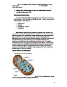
Nucleus:
Genetic material is DNA (deoxyribonucleic acid). It is usually found in every cell, from microorganisms that have only one cell, to plants and animals that have many cells, where the cells make up tissues and organs. The cell and its constituents can be seen only with the help of increasingly powerful microscopes (see Fig.1).
Source:
Inside a cell from a plant or animal, the genetic material is enclosed in a spherical compartment, the nucleus. It is packaged into long compact structures called chromosomes. The totality of all the genetic material packaged into chromosomes is the genome. Each species has a different genome. For example, there are 23 pairs of chromosomes in the human genome, one of each pair from each parent. Bacteria have chromosomes which are not enclosed in a nucleus. The E.coli bacterium, which lives in the gut of mammals and human beings, has only one chromosome in its genome ().
A basic way of describing a chromosome is a long molecule of DNA which is wound up and attached around special proteins this forms chromatin. In animals and plants each chromosome is doubled but remains joined at one point. When the DNA molecule is stripped of bound proteins it consists of two strands which are joined together in a double helix. Each individual thread is joined up end to end by a long string of units. There are four individual units of DNA they are labelled A, T, G and C, which stand for the identifying bases for each unit, adenine, thymine, guanine and cytosine.
Nucleus is the “brain” of the cell and is contained within the nuclear membrane (which insures that the interior of the nucleus is isolated from the cells cytoplasm allowing two different environments to be maintained) it contains pairs of chromosomes each carrying hereditary DNA (Toole, G & S: 1984) the nucleus controls every organelle with the Cytoplasm.
Within the nucleus is the Nucleolus, It is a small structure within the nucleus that forms ribosome’s which then pass into the cytoplasm to produce protein (Moth.E: 2003). The cell membranes is a fine membrane made of protein and lipids, it has two functions to keep the cytoplasm and the nucleus in.
Endoplasmic Reticulum:
The endoplasmic reticulum is found in the cytoplasm of the cell and like the plasma membrane is composed of phospholipids and protein. ER is an intracellular transport system moving materials from one part of the cell to another. In certain parts of the cell the ER is continuous with the plasma and nuclear membranes providing a route by which materials might move from the nucleus to the cytoplasm. There are two types of ER, rough and smooth. On the cytosol side of the ER membrane there are ribosome’s attached to it and this is known as Rough ER. This forms the bulk of the ER and consists of flattened cavities, its function is to isolate and transport the proteins that have been synthesised by the ribosomes.
Here is a diagram of ER;
Source:
The major function of ribosomes is protein synthesis; RER allows the conveyance of the finished product via other organelles to the plasma membrane for export. Smooth ER consists of tubular cavities and does not have ribosomes attached, it is concerned with synthesis and transport of lipids and steroids. Proteins produced by the ribosomes are believed to move to the Golgi apparatus via Rough ER
The Golgi apparatus is an assembly point through which raw materials for secretion are funnelled before being shed from the cell. It consists of a stack of flattened membrane bound sacs continuously being formed at one end and budded off as vesicles at the other. At times the Golgi modifies the cell products it contains; the protein received from the RER has polysaccharides added to it producing glycoproteins. Similarly it modifies lipids into Glycolipids; these products are then wrapped into various types of secretary vesicles for movement to the cell membrane so they can discharge their contents. After which the membranes of the vesicles become fused with the plasma membrane thereby providing the cell with additional surface area. The vesicles also contain digestive enzymes and become Liposome’s.
Ribosomes
Ribosome’s are cytoplasmic organelles discovered in both prokaryotic and eukaryotic organisms. Found in great abundance up to 10,000 in bacterial cells and many times more in eukaryotic cells, they comprise of proteins and rRNA molecules known as subunits, to form a large ribosomal complex. Both eukaryotic and prokaryotic ribosome’s in association with transfer RNA (tRNA), act as a site for mRNA translation, assembling a specific sequence of amino acids into polypeptide chains, once the mRNA joins the two component subunits (large and small) of the ribosome. The tRNA is covalently bonded to an individual amino acid and has a complimentary nucleotide sequence, an anticodon, to each mRNA codon which forms base pairs, adding specificity to the selection of the corresponding amino acids. The mRNA is linked by hydrogen bonds to the tRNA and is held in proximity to the amino acid so that a peptide bond is formed, this process occurs again and each amino acid is polymerized into a growing peptide chain.
Here is a diagram of a ribosome:
Source:
Ribosome’s existed in two distinct forms; free and bound and may be positioned in several locations throughout the cell depending on cell function. Free ribosome’s can occur individually, a monosome, or in clusters called polyribosome’s or polysomes and are found in the cytosol (the fluid component of cytoplasm, excluding organelles and the insoluble, usually suspended, cytoplasmic components). Found in greater concentrations in cells that retain proteins, they manufacture proteins that are either held in solution in the cytoplasm or those used in the formation of cytoplasmic structural and motile elements. Bound ribosome’s are situated on the outside of the endoplasmic reticulum[1] forming rough ER (RER) and a large number are seen in cells that make proteins to be secreted out of the cell, such as pancreatic cells producing digestive proteins. The bound ribosome’s form proteins that may be utilised for the cell membrane, packaged into vesicles for storage, as well as those required for export from the cell ().
Ribosomes are also located in mitochondria and chloroplasts within eukaryotic organisms, these are smaller than those found in the cytoplasm and can be compared to bacterial ribosomes.