Introduction
The cell surface membrane (formerly called the plasma membrane) surrounds the cytoplasm of eukaryotic cells. The membrane forms a selectively permeable barrier, controlling the substances that enter and leave the cell and therefore enables the cell to regulate its internal environment.
Structure
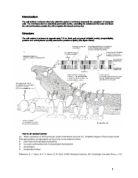
The cell surface membrane is approximately 7.5 nm thick and composed of lipids (mainly phospholipids), proteins and carbohydrates (usually attached to proteins or lipids). (See figure below).
THE FLUID MOSAIC MODEL
In 1972, Singer and Nicolson put forward the 'fluid mosaic' model of membrane structure in which protein molecules float about in a fluid phospholipid bilayer. The scattered protein molecules resemble a mosaic but, since the phospholipid bilayer is fluid, the proteins form a fluid mosaic pattern. Why fluid mosaic?
Fluid - because the protein molecules float about in a fluid phospholipid bilayer. The membrane is held together mainly by hydrophobic interactions between the phospholipids and between proteins and phospholipids. These weak interactions allow the molecule to move. Phospholipid molecules move in the plane of the membrane. Proteins are much larger and move more slowly - imagine protein molecules moving about like icebergs in a 'sea' of lipid.
Mosaic - because the membrane is made of different types of molecules arranged in a mosaic pattern. A membrane is like a collection of many different proteins, cholesterols, glycoproteins and glycolipids in the phospholipid bilayer.
The membrane has the following features:
* It is between 7 to 9 nm thick.
* The basic structure is a phospholipid bilayer.
* The hydrophilic phosphate heads of the phospholipids face onwards into the aqueous environment inside and outside the cell.
* The carbohydrate tails face inwards and create a hydrophobic interior.
* The phospholipids are fluid and move about rapidly by diffusion in their own layers.
* Some of the fatty acid tails are saturated and some are unsaturated. Unsaturated tails are bent and fit together more loosely. Therefore the more unsaturated the tails are, the more fluid the membrane is.
* Most protein molecules float about in the phospholipid bilayer forming a fluid mosaic pattern.
* The proteins stay in the membrane because they have regions of hydrophobic amino acids which interact with the fatty acid tails to exclude water. The rest of the protein is hydrophilic and faces into the cell or out into the external environment, both of which are aqueous.
* Some proteins penetrate only part of the way into the membrane while others penetrate all the way through.
* Some protein and lipids have short branching carbohydrate chains like antennae, forming glycoproteins and glycolipids and keeps them more fluid. This can be important for organisms living at low temperatures when membrane can solidify. Cholesterol also increases flexibility and stability of membranes. Without it, membranes break up.
* The two sides of a membrane can differ in composition and function.
LIPIDS
There are three types of lipid in the cell surface membrane:
. Phospholipids - which make up 75% of the lipid. Phospholipids consist of a glycerol molecule plus two molecules of fatty acid and a phosphate group. The phosphate / glycerol head is hydrophilic (it attracts water) and the fatty acid tails are hydrophobic (they repel water).
In aqueous (water-based) solutions phospholipids automatically arrange themselves into a double layer so that the hydrophobic tails pack together inside the layer ...
This is a preview of the whole essay
* The two sides of a membrane can differ in composition and function.
LIPIDS
There are three types of lipid in the cell surface membrane:
. Phospholipids - which make up 75% of the lipid. Phospholipids consist of a glycerol molecule plus two molecules of fatty acid and a phosphate group. The phosphate / glycerol head is hydrophilic (it attracts water) and the fatty acid tails are hydrophobic (they repel water).
In aqueous (water-based) solutions phospholipids automatically arrange themselves into a double layer so that the hydrophobic tails pack together inside the layer away from the water, and the
The phospholipid bilayer forms spontaneously with the non-polar fatty acid chains facing inwards towards each other and the polar phosphate groups facing outwards into the extra-cellular fluid and the inside of the cell (both of which are water-based environment).
The interaction between the hydrophobic and hydrophilic ends helps give the membrane stability and it is also these lipids which give the membrane selective permeability. Lipid soluble (hydrophobic) molecules easily pass through the membrane by diffusion whilst hydrophilic substances cannot diffuse through; instead they cross the membrane via water-filled pores or channels in intrinsic proteins.
2. Glycolipids - which make up 5% of membrane lipids. Glycolipids are lipids that have combined with polysaccharides. They are found in the outer layer of cell membranes and the carbohydrate portion of the glycolipid extends into the intercellular space and is called a glycocalyx. These are important in cell-cell recognition.
3. Cholesterol - belongs to a group of lipids called steroids. It is present in all cell membranes except those of bacteria. It can make up to 25% of the lipids in animal cell membranes but isn't found so much in plant membranes. Having cholesterol molecules between phospholipid molecules makes the membrane less fluid and more stable.
All lipids can move sideways (laterally) within the membrane and exchange position with each other. This gives the membrane fluidity which is essential in processes such as phagocytosis. The degree of fluidity of call surface membranes is determined by:
. The length of the fatty acid side chains (the longer the chains, the lower the fluidity).
2. The proportion of the fatty acids which are saturated (the higher the percentage of saturated fats, the lower the fluidity).
3. The steroid content (the higher the steroid content, the lower the fluidity).
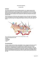
PROTEINS
Intrinsic proteins - those which are tightly embedded in the membrane, and may extend from one side of the membrane to the other. They are usually glycoproteins. They have five main functions:
. Channel proteins - form a tiny gap in the membrane to allow water soluble molecules and ions through by diffusion.
2. Carrier proteins - carry water soluble molecules and ions through the membrane by active transport and facilitated diffusion.
3. Receptor proteins (transmembrane proteins). Recognise and bind to specific molecules (e.g. hormones). Proteins have very specific shapes. This makes them ideal as receptor molecules for chemical signalling between cells. For example, hormones are chemical messengers which circulate in the blood but only bind to Neurotransmitters, the chemicals which enable nerve impulses to pass from one nerve cell to the next, also fit into specific receptor proteins in nerve cells.
4. Enzymes can be embedded in the inner membrane of a cell or organelle Proteins sometimes act as enzymes, for example the microvilli on epithelial cells lining some parts of the gut contain digestive enzymes in their cell surface membranes. For example, ATPase.
5. Proteins in membrane also help strengthen the membrane. There are hydrogen bonds between the protein and the hydrophilic heads of the phospholipids.
As in lipids, intrinsic proteins have a hydrophobic and hydrophilic region and the interaction between these regions confers stability on the membrane.
Glycoproteins - (also found in the outer layer) are proteins with attached polysaccharides of short, branched chains of monosaccharides. Glycoproteins have a variety of specific shapes due to the different branching patterns of the monosaccharides. These allow different cells to recognise each other. For example, some glycoproteins are antigens - they are recognised by white blood cells, which start an immune respond.
Extrinsic proteins - those which are entirely outside of the membrane (lying on the surface of membrane), but are bound to it by weak molecular attractions (ionic , hydrogen , and/or Van der Waals bonds) and many have a carbohydrate (glycocalyx) which extends into the intercellular space.
Functions
Cells and many of the organelles inside them are surrounded by membranes, which have a range of functions:
Membranes around organelles:
. Divide the cells up into different compartments to make the different functions more efficient e.g. the substances needed for respiration (like enzymes) are kept together inside mitochondria.
2. Provide a big surface area for enzymes or pigments that help the organelle does its job, e.g. chlorophyll pigment in chloroplast (needed for photosynthesis).
3. Protective - e.g. the membrane of lyosomes keeps hydrolytic (digestive) enzymes apart from the cell contents which they would otherwise destroy.
4. Can be used to form vesicles to transport substances between different areas of the cell.
Membranes around cells:
. Regulate transport in and out of cell. (see "Transport across the cell surface membrane")
2. Allow cell recognition - allow recognition by other cells, e.g. the cells of the immune system
3. Provide receptors for molecules like hormones.
4. Act as a barrier to many substances.
5. Are permeable to small molecules like oxygen and water, and are selectively permeable to ions and larger molecules e.g. glucose.
6. Form specialized junctions that allow cell adhesion or communication.
Transport across the cell surface membrane
Substances may move across the membrane by:
. Diffusion
2. Osmosis
3. Facilitated diffusion
4. Active Transport
The table below shows a summary of methods of transport across membranes.
Diffusion
Facilitated diffusion
Osmosis
Active transport
Definition
Movement from high to low concentration using kinetic energy of the molecules involved.
Movement from high to low concentration involving carrier protein in the cell membrane.
Movement of water from dilute solution to a more concentrated one using kinetic energy of the water molecules.
Movement from low to high concentration using energy supplied by cellular ATP.
Type of molecules
Small enough to pass between phospholipids in bilayer, or larger and lipid soluble
Carry a charge, or are not lipid soluble. If water soluble they may pass through channels that contain water
Water molecules
Ions that are moving from an area of high to an area of low concentration
Examples of molecules
Vitamin D,
CO2, O2
Glucose
Water molecules
sodium (Na+) potassium (K+) chloride (Cl-) hydrogen (H+)
ATP needed
No
No
No
Yes
Direction of movement
Down the concentration gradient.
Down the concentration gradient.
Down the water potential gradient.
Against the concentration gradient.
Proteins in membrane involved
No
Protein channels or pores, which have the hydrophilic groups of the amino acid side chains lining the inside of them so that the channel is filled with water. Water soluble molecules can pass through. May use protein carriers. Some channels are open all the time and some open in response to attachment by a hormone molecule.
May or may not.
In collecting ducts of kidney tubules water molecules pass through membranes by osmosis, through water channels, opened by action of the ADH (anti diuretic hormone).
Yes - carrier proteins or protein pumps
Abnormalities
CYSTIC FIBROSIS
A genetic disease causes an abnormality in the mucus normally found in the lungs, resulting in increased bacterial infections and difficulty breathing.
Cystic fibrosis is caused by a mutation in a gene called the cystic fibrosis transmembrane conductance regulator (CFTR). The product of this gene helps create sweat, digestive juices, and mucus.
Cystic fibrosis occurs when there is a mutation in the CFTR gene. The protein created by this gene is anchored to the outer membrane of cells in the sweat glands, lung, pancreas, and other affected organs. The protein spans this membrane and acts as a channel connecting the inner part of the cell (cytoplasm) to the surrounding fluid. This channel is primarily responsible for controlling the movement of chloride from outside the cell into the cell. When the CFTR protein does not work, chloride is trapped outside the cell. Because chloride is negatively charged, positively charged ions also cannot cross into the cell because they are affected by the electrical attraction of the chloride ions. Sodium is the most common ion in the extracellular space and the combination of sodium and chloride creates the salt which is lost in high amounts in the sweat of individuals with CF. This lost salt forms the basis for the sweat test.
DIABETES MELLITUS
Some human diseases are the result of faulty membrane transport systems. An example would be type II (adult onset) diabetes mellitus. Excess glucose in the bloodstream, caused by eating a meal rich in carbohydrates, is usually taken up by myocytes (muscle cells) and adipocytes (fat cells). The glucose transporter GluT4 is normally present in the cell membrane in small amounts. The presence of insulin (a hormone secreted by the pancreas in response to high glucose levels) causes more GluT4 transporters to be exposed, increasing uptake of glucose into the cell. In type II diabetes there is resistance to the metabolic effects of insulin, either at the cell membrane or in post-receptor signaling systems. This means that little glucose can be taken up by myocytes and adipocytes, and high blood glucose levels are the result.
Conclusion
The biological membrane is a collage of many different proteins embedded in the fluid matrix of the lipid bilayer. The lipid bilayer is the main fabric of the membrane, and its structure creates a semi-permeable membrane. The hydrophobic core impedes the transport of hydrophilic structures, such as ions and polar molecules but enable hydrophobic molecules, which can dissolve in the membrane, cross it with ease. Proteins determine most of the membrane's specific functions. The plasma membrane and the membranes of the various organelles each have unique collections of proteins. For example, to date more than 50 kinds of proteins have been found in the plasma membrane of red blood cells.
NUMBER OF WORDS: 2130
Bibliography
Books
* D. J. Taylor, N. P. O. Green, G. W. Stout. (2006) Biological Sciences 1&2, Cambridge University Press, ISBN 0-521-56178-7
* Dennis L. Kasper MD, Anthony S. Fauci MD, Dan L. Longo MD, Eugene Braunwald MD, Stephen L. Hauser MD, J. Larry Jameson MD, PhD (2004) Harrison's Principles of Internal Medicine 16th Edition, McGraw-Hill Professional; 16 edition, ISBN: 0071402357
* Richard Fosbery (2002) AS Biology, philip allan updates, ISBN 0-86003-670-7
Internet
* Healthline, Available from:
http://www.healthline.com/adamcontent/cystic-fibrosis [Accessed 6th December 2006]
http://www.healthline.com/adamcontent/diabetes [Accessed 6th December 2006]
* University of Texas Medical Branch (2003) Cell Biology Graduate Program
Texas, University of Texas, Available from:
http://cellbio.utmb.edu/cellbio/membrane_intro.htm [Accessed 6th December 2006]