The structure and function of chloroplasts
The plant anatomist N. Grew first saw chloroplasts in the 17th century with a light microscope. They were found to be unique to green plant cells, (also certain protists), but their role was not understood until the late 19th century, (Sadava, David.E. 1993).
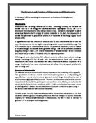
Diagram of a Chloroplast
http://www.cbs.dtu.dk
The name chloroplast originates from the Greek words chloros and plastos, meaning green and moulded. Chloroplasts contain the green pigment chlorophyll, which absorbs red, blue and violet light and reflects green light giving the plant an overall green colour. Chlorophyll absorbs so strongly that it can mask other less intense colours. Some of theses more delicate colours, from molecules such as carotene and quercetin, are revealed when the chlorophyll molecules decay in autumn and the woodlands turn red, orange and golden brown. Chlorophyll plays a central role within the plant as it consumes light energy and initiates the event of photosynthesis. The primary function of chloroplasts is photosynthesis, a process where the organelle converts carbon dioxide and water into glucose and oxygen using solar energy. A mature plant leaf cell usually contains twenty to one hundred chloroplasts although the number may vary significantly depending on the species and location. Chloroplasts have a tendency to be shaped like flattened spheres, but again the exact size and structure is relative to the function. Sizes of these photosynthetic organelles range between 4 to 6 microns in diameter and 1 to 5 microns in length, (Mader, Sylvia.S. 2001). They are found in the cytoplasm of the plant cell and belong to the group of organelles known as the plastids. Among the plastids are also the amyloplasts, which store starch and the chromoplasts that contain red and orange pigments. An envelope of two phospholipid membranes, the outer membrane and the inner membrane surround each chloroplast. The two membranes are separated by a narrow intermembrane space. The outer membrane is quite rigid in structure and contains porins that make it permeable to small organic molecules and ions. The inner membrane acts as a barrier as transport proteins prevent anything other than water, carbon dioxide and oxygen crossing into the internal cavity. The inner part of the chloroplast is named the stroma and is a gel like matrix containing many enzymes, starch grains, some pigments, protein, ribosomes and the circular chloroplast DNA. The stroma is the location where light independent reactions occur and also contains the enzymes required for the Calvin cycle. Chloroplasts also feature a third internal membrane system, the thylakoids. Thylakoids are flat sac like structures suspended in the stroma and are usually arranged into stacks termed grana. The area inside the internal membrane system is referred to as the thylakoid space and contains the pigment chlorophyll. The extensive folding of the inner membrane provides a great increase in surface area, making it approximately 600 times larger. The membranes of the grana hold the electron carriers needed for light dependant reactions, ATP synthetase, and make synthesis possible. The internal layer of the membrane is hydrophobic so the hydrophilic heads of the chlorophyll molecules lie flat on the surface, not unlike solar panels. The chlorophyll molecule also has a hydrophobic tail end which projects into the thylakoid membrane and acts like an anchor.
The differences between mitochondria and chloroplasts
Chloroplasts are only found within phototrophs, (plants and some algae), whereas mitochondria are found inside chemotrophs, (both plants and animals). The gas oxygen is produced by chloroplasts and carbon dioxide gas is given off by mitochondrion. Chloroplasts, unlike mitochondria, may use chemical energy or solar energy to initiate the process of photosynthesis. The purpose of photosynthesis is to create glucose and some oxygen from the three components sunlight, water and carbon dioxide. The glucose can then be processed by the mitochondria in to, carbon dioxide, water and the universal energy currency, ATP. The energy of ATP is used for active transport, synthetic reactions and all energy requiring processes in the cell, (Jones, Mary., Gregory, Jennifer. 1999). When mitochondria convert glucose into ATP the action is referred to as cellular respiration, chloroplasts do not respire. Mitochondria perform aerobic respiration in four main stages, glycolysis, the link reaction, the Krebs cycle and finally the electron transport chain, also named oxidative phosphorylation. For each molecule of glucose entering respiration, two molecules of ATP are produced from glycolysis, two from the Krebs cycle, and thirty-four from oxidative phosphorylation. Therefore, one molecule of glucose can form thirty-six to thirty eight molecules of ATP energy. Mitochondria and chloroplasts are structurally different; chloroplasts tend to be shaped irregularly and are larger. Internally mitochondria possess cristae, F0F1 complexes and a matrix region unlike plant organelles. The chloroplast contains a third thalkaloid membrane, a stroma and grana unlike the mitochondria. The two organelles store and use different enzymes and only plant cells appear green due to the amount of chlorophyll housed within the chloroplast. Only chloroplasts have the ability to photosynthesise, a process that also occurs in stages. Firstly there are the light dependant reactions photoreduction and phosphosphorilation. During photoreduction electrons donated form the water generate NADPH, (nicotinamide adenine dinucleotice phosphate, plus electron). Photophosphorilation involves the use of a proton gradient to manufacture ATP. The products of theses two light dependant reactions are then fed into the Calvin cycle, a dark reaction. The Calvin cycle acts to fixate and reduce carbon dioxide and regenerate RuBP, (ribolose bisphosphate. RuBP is a five-carbon molecule needed to keep the Calvin cycle turning. For every three turns of the Calvin cycle one molecule of PGAL is generated, (Becker, Wayne.M., et al. 2000). Glyceraldehyde-3-phosphate or PGAL can be converted into starch and glucose and then passed to the mitochondria to produce ATP energy. Scientists theorise that mitochondria may have evolved from ancient purple bacteria and state that they may only be inherited from the female parent. Chloroplasts are thought to have originated from ancient cyanobacteria and can be inherited form either parent.
The similarities between mitochondria and chloroplasts
Both mitochondria and chloroplasts were discovered around the same time historically. The two organelles are both located predominantly in the cells that require the most energy and are situated within the cytoplasm. An envelope of two phospholipid membranes surrounds mitochondria and chloroplasts. The outer membranes are both covered with porins that permit selective molecules to cross, while the inner membranes act as more of a barrier. Chloroplasts and mitochondria have an intermembrane space and contain intricately folded internal membranes that serve to increase surface area. Both organelles can produce some of their own proteins and have their own ribosomes and DNA, allowing them to divide independently within the cell by the process of binary fission. The major similarity between mitochondrion and chloroplasts is that functionally they work to convert simple molecules in to chemical energy by making and braking bonds, and are both concerned with ATP. Mitochondria and chloroplasts can be described as being semiautonomous organelles, as they contain their own genetic material in the form of circular DNA, mRNA, tRNA and ribosomes, (Becker, Wayne.M., et al. 2000). These features led biologists to formulate the endosymbiont theory. It was suggested that approximately one and a half billion years ago they were both separately engulfed through phagacytosis by larger membranous organisms. The relationship between the bacterium and the hosts became mutually beneficial and symbiosis was established. As the host cells and cytoplasmic bacterium adapted to living together over hundreds of millions of years the bacterium changed some of their functions and developed into either mitochondria or chloroplasts.
Conclusion
Chloroplasts are extremely important organelles as they are the sites of photosynthetic reactions. Photosynthesis is either directly or indirectly crucial to almost all life on earth. During the process carbon dioxide is removed from the atmosphere and oxygen gas is produced. Oxygen is vital to all aerobic forms of life. Photosynthesis within chloroplasts provides plants with energy allowing them to grow, reproduce and to be utilised as food by other organisms. Mitochondria are essential for cellular respiration within both plants and animals. They require oxygen to complete the process and produce carbon dioxide as a waste product. Both mitochondria and chloroplasts have structural differences but are integrally linked by their functions The excess carbon dioxide emitted into the atmosphere via respiration is constantly being reabsorbed by the process of photosynthesis. While the excess oxygen produced by photosynthesis is removed during the continual event of cellular respiration. The physical structure of both organelles is inversely related to the location and energy requirements of the cell. To conclude, mitochondria and chloroplasts have opposite yet similar functions. They both work to produce energy for the organism but using different methods. Chloroplasts require carbon dioxide and expel oxygen whereas mitochondria need oxygen and produce carbon dioxide. The different, opposite actions of the two organelles function to maintain a mutually beneficial relationship. This harmonious relationship between chloroplast and mitochondria also benefits and supports almost every form of life on our planet.
Reference list
Sadava, David.E. 1993. Cell biology, organelle structure and function. Jones and Bartlett Publishers International. Pages 2-4, 92-124, 128-200.
Mader, Sylvia.S. 2001. Biology. 7th edition. McGraw-Hill companies, Inc. Pages 60-64, 70-71, 112-113, 128-140.
Taylor, D.J, et al. 2000. Biological Science 1 & 2. 3rd edition. Cambridge University Press. Pages 128-140, 196-215, 264-278.
Wilson, Kathleen.J.W., and Waugh, Anne. 1996. Ross and Wilson Anatomy and Physiology in Health and Illness. 8th edition. Churchill Livingstone. Pages 18-20.
Tortora, G.J., and Grabowski, S.R.2000. Principles of Anatomy and Physiology. 9th edition. John Wiley and Sons Inc. Pages 60-62, 84-87.
Hurry, Stephen.W. 1975. The Microstructure of Cells. Williams Clowes and Sons Ltd. Pages 19-24, 44-48.
Becker, Wayne.M., Kleinsmith, Lewis.J., Hardin, Jeff. 2000. The world of the cell. 4th edition. Addison Wesley Longman Inc. Pages 88-91, 405-484.
Jones, Mary., Gregory, Jennifer. 1999. Central Concepts in Biology. Cambridge University Press. Pages 1-21.
http://www.cbs.dtu.dk/dave/roanoke/bio101ch06.htm (Biology 101 lecture notes, Roanoke College, Virginia).