TABLE 5.1 Thoracic organs. Rat circulatory system organs being indentified
- Photos of the full dissection and organs were taken for records and drawing purposes.
- The heart was examined, sketched and labeled accordingly.
b) Frog dissection and transport system
- A freshly anesthetized frog was neatly pinned onto the dissection board with its ventral side facing upwards.
- The upper epidermal layer was cut upwards from in-between the hind legs towards the jaw of the frog with a 1-blunt sided scissors.
- The area below the upper epidermal layer was spaced using a seeker/probe.
- The skin was cut and removed using a scalpel blade.
- The skin/upper epidermal layer was pinned onto the dissection board.
- The anterior abdominal vein that would be found along the midline of the frog, slightly below the ventral abdominal wall was observed. The vein was pinpointed about halfway in between the pelvic curve and the sternum.
- Two small incisions of about 0.5cm long were made on the left and right of the pinpointed area using a pair of scissors.
- A knot to the top and bottom of this incision was made to tie the pinpointed area of the abdominal anterior vein.
- A horizontal cut was made in between the knots made on the vein.
- The abdominal muscle wall was cut upwards/vertically on both sides of the anterior abdominal vein. A straight line was maintained all the way while blood vessels below or the organs were ensured not to be pierced.
- The tissue strip and the bone that was cut were both lifted. They were then released and removed from the organs and the anterior abdominal vein.
- In a similar manner to step 11, vertical cuts and horizontal cuts were made as the figure below and this layer was pinned onto the board. The visceral organs in the abdominal cavity were revealed.
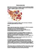
- The thin layer called the pericardium surrounding the heart was removed with the help of a pair of scissors and forceps.
- Photos of the heart were taken for records and drawing purposes. The diagrams were labeled accordingly.
TABLE 5.1 Thoracic organs. Frog circulatory system organs being indentified
3.0 Results
a) Rat transport system
4.0 Discussion
Comparing the anatomy and physiology of both transport systems (Mammalian and Amphibian)
TABLE 5.3 Anatomy and physiology. The comparison of transport systems in
mammals and amphibians
Based on the observation of the table above, the septum is a key evolutionary adaption that is present in the mammalian heart but not in the amphibian. Its development ensures the complete separation of oxygen-rich and poor blood as seen in the mammal. This is a more effective form of transport and exchange because waste materials carried by oxygen-poor blood do not interact with the oxygen-rich blood that gets delivered to the rest of the body. As endotherms, mammalians generate their own thermal energy to regulate internal temperature. Hence, a higher metabolic rate is needed and their circulatory system is more complex.
Gaseous exchange in frogs is more effective than in rats. Frogs are amphibians that have a thin, moist skin that is very important in the exchanging of gaseous materials. This form of respiration is beneficial because the higher surface area on the skin allows gaseous exchange and the dissolving of those gases.
Rats are more effective in the transport of solutes and gases because of the presence of the septum mentioned earlier.
5.0 Conclusion
Rats are more evolutionarily advanced compared to frogs because of the development of the septum in the heart that ensures no mixing of substances.
6.0 Answers to questions
- What is the largest blood vessel in the amphibian and the mammal?
The aorta is the largest blood vessel in the amphibian and the mammal.
- Explain the difference between different blood vessels.
Blood vessels consist of arteries, capillaries and veins. The table below will compare their differences.
TABLE 5.3 Blood vessels. Comparison of different blood vessels.
Arteries are muscular blood vessels involved in the transport of oxygenated blood away from the heart (in exception to the pulmonary artery). Arteries carry oxygen-rich blood at a relatively high pressure; hence they are tougher and thicker than the other vessels. They branch off into smaller arterioles that channels oxygen-rich blood to the capillaries.
Capillaries are very thin blood vessels that deliver oxygen-rich blood from arteries. Their thin walls allow the exchange oxygen and carbon dioxide into the surrounding tissues. The hemoglobin in the capillaries will supply the oxygen. Then tissue releases its waste products, e.g. carbon dioxide, into the surrounding erythrocytes. These erythrocytes will carry the now waste-rich blood to the veins which is transported back to the heart and lungs through the veins.
Veins are blood vessels that channel waste-rich blood back to the heart and lungs from smaller venules. Veins are not as tough compared to arteries because blood is transported at a lower pressure. They have valves called sphincters inside them, which assist in ensuring a one direction blood flow. This characteristic is present in the vein due to blood flowing against the force of gravity.
- What is the point in cutting the anterior abdominal vein in the frog?
The anterior abdominal vein is tied and cut to avoid hemorrhaging major arteries and veins. It is performed to cease the blood flow to the heart that might cause leakage of blood into the abdominal cavity.
- Which animal bled less when cut? Why?
The rat bled less when cut. The blood composition of rats (mammal) has platelets that contribute to clotting factors. Platelets in frogs however, do not have platelets but fibrinogen. Also, the frog has more blood vessels near the skin to allow exchange of gases and cutting its skin layer might cause some rupturing of those vessels.
- Give details of the main anatomical differences of the transport systems that you observed between the mammal and amphibian.
The difference seen in the transport systems in the mammal and amphibian is the number of chambers in the heart. The mammalian heart has four chambers consisting of 2 atria and 2 ventricles. The amphibian heart has only three chambers consisting of 2 atria and 1 single ventricle.
The difference here lies in the presence of the septum that divides the ventricle region. The mammalian heart has a septum while the amphibian heart does not.
Without the presence of the septum, there is a mixing of oxygen-rich blood and oxygen-poor blood within the amphibian heart. In the mammalian heart however, there is a complete separation of the two types of blood and no mixing of the both.
- Explain how essential molecules (i.e. oxygen) get transported in an amphibian.
The skin of the frog is an essential tool for respiration. Its large surface area allows optimum exchange and dissolving of gases. This type of respiration is called cutaneous respiration. The skin of the frog consists of blood capillaries and has cutaneous glands that secrete mucus. The mucus retains the skin’s moisture and maintains a thin film of water underneath the skin’s surface. This enables the exchange of oxygen through active transport between the blood vessels and the environment.
- Make academically sound inference(s) as to why there is a noted difference between an amphibian and a mammal.
A difference is observed due to the presence of a septum in a mammalian heart. Amphibians have a three chambered heart: 2 atria and 1 ventricle. This single ventricle is not separated by a septum; unlike the 2 ventricles in the mammalian heart that includes a septum. Without the septum, the exchanging of materials are not as effecting because there is a degree of mixing between oxygen-rich blood and oxygen-poor blood that carries waste materials. This can cause the waste materials to be circulated back into the body.
In a mammalian heart, a full septum is seen that divides the ventricles into two. It has a more effective transport and exchange of oxygen-rich and oxygen-poor blood to organs that need it. The presence of a septum in the heart proves to make the mammal more adaptive to evolution.
7.0 Format and referencing
-
Prater, A. M., Sep 24, 2009. The Different Blood Vessels: Arteries, Veins and Capillaries [Online]. Available from: [Accessed 11 November 2010]
-
Ross, J., June 30, 1999. Blood Vessels [Online]. Available from: [Accessed 11 November 2010]
-
Simmons, K., 2006. Rat circulatory system [Online]. University of Winnipeg. Available from: [Accessed 14 November 2010]