2NADH + O2 + 2H+ 2NAD+ + 2H2O ΔEo=1.14V
Which can be broken down into the following half-equations:
NADH NAD+ + H+ + 2e-
O2 + 4H+ + 4e- 2H2O
The potential difference between NADH and oxygen is very high (1.14V) because the electrons in NADH (and FADH2) have a high transfer potential, whilst oxygen has a high affinity for electrons (a high redox potential shows that the reaction is heavily oxidising). This produces a large thermodynamic driving force, which drives the movement of electrons through the electron transport chain (ETC) (Stryer (1995)). As the electrons move through the ETC they are used to reduce successive electron carriers, which are oxidised when they move on (redox reactions), until they are passed on to oxygen. The electron carriers are redox pairs, like NADH and NAD+; for example cytochrome b contains iron at different oxidation states. As the electrons reduce and then oxidise successive electron carriers free energy is released in a series of discrete steps. The liberation of free energy is therefore controlled so that it is produced in small quantities sufficient to drive the proton pumps, which set up the electrochemical gradient. If the liberation of free energy were not controlled in this way then it would be produced all at one time, mainly as heat, and so the process would be very inefficient. The energetics of electron transfer (the redox potentials of the electron carriers) depends on the microenvironment (where the ions are); hence the same redox couple can produce different amounts of free energy in different microenvironments (e.g. Fe3+/Fe2+). Therefore, different numbers of protons can be pumped across the membrane by ETC in different environments (e.g. in mitochondria or chloroplasts). Eventually low energy electrons are passed back to H+ and are used to reduce oxygen to water.
In this manner, the electron transfer potential of compounds like NADH is converted into the proton motive force, which drives ATP synthesis, storing the electron-motive force in the phosphoryl potential in ATP.
Mitochondria:
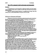
In mitochondria the ETC is located in the inner mitochondrial membrane and it pumps protons from the mitochondrial matrix into the intermembrane space between the inner and outer mitochondrial membranes. These therefore are the two cellular compartments mentioned above. This creates an electrochemical gradient between the intermembrane space and the mitochondrial matrix, which drives ATP synthesis. ATP synthase and the mitochondrial ETC are located on the inner membrane, which is highly folded into a series of ridges called cristae to increase the surface area. Figure 1 shows a mitochondrial ETC. It consists of three protein complexes – complex I is the dehydrogenase complex, III the b-c1 complex and IV the cytochrome oxidase complex. Each contains multiple oxidation-reduction centres. In I the main redox couple is NAD+/NADH, in III it is Fe3+/Fe2+, and in IV it is Cu2+/Cu+. Ubiquinone transfers electrons from complex I to complex III and cytochrome c shuttles electrons between III and IV. Both of these are small molecules that can diffuse rapidly within the inner mitochondrial membrane. Some complexes are very large, for example NADH dehydrogenase consists of 22 polypeptides in a molecule 800,000 daltons in size. The complexes are not fixed in the mitochondrial membranes but are free to diffuse – this gives sufficient contacts to allow the electron transport process to take place. As an electron moves through a redox couple a proton is pumped across the mitochondrial membrane, because the change in redox state presumably causes a conformational change, which transfers the proton. Complexes I and III transfer four protons for every two electrons, whilst IV only transfers two protons. Therefore the transport of two electrons down the ETC transfers ten protons from one side of the membrane to the other. Complex II is the FADH2 dehydrogenase complex, from which electrons are passed on to complex IV by ubiquinone but no protons are pumped across the membrane. One ATP molecule is produced for every four protons that move across the membrane down the electrochemical gradient, that is, 2.5 ATPs are produced for every two electrons that pass down the electron transfer chain. This, however, is not the entire story because NADH produced by glycolysis in the cytosol cannot be transported directly into the mitochondrial matrix: it must be brought in indirectly by a ‘shuttle’ system that transfers another reduced compound into the mitochondria; this consumes ATP.
NADH and FADH2 therefore provide the electrons that power the electron transport chain and are oxidised in the process (to NAD+ and FAD+). If the electron transfer chain is to continue they must be regenerated from their oxidised forms by oxidising fatty acid and carbohydrate stores. The principal pathways are –
-
Glycolysis – the conversion of carbohydrate to pyruvate reduces NAD+ to NADH.
-
β-oxidation of fatty acids to acetyl-CoA generates NADH and FADH2.
-
The TCA cycle – oxidation of acetyl-CoA to carbon dioxide generates NADH and FADH2.
Fatty acids contain a more reduced form of carbon than carbohydrates and so the complete oxidation of a fatty acid to carbon dioxide and water generates more reduced coenzyme and therefore more ATP than a comparable carbohydrate. For example, the maximum possible yield of ATP is 104 molecules from the oxidation of one molecule of palmitate and 31 molecules from the oxidation of one molecule of glucose. The process is about 40% efficient – i.e. 40% of the available free energy can be stored in the ATP molecules during oxidative phosphorylation
Chloroplasts:
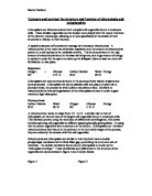
The reduced carbon energy source that is oxidised by the mitochondria (such as carbohydrate) is synthesised in the chloroplasts of plants by reductive biosynthesis, using sunlight to drive the process (hence it is called photosynthesis). ATP is required by reductive biosynthesis as well, but it is synthesised by photophosphorylation – using sunlight, as the name suggests, as a source of free energy rather than the oxidation of complex molecules. Figure 2 shows the reactions of photosynthesis.
Light is captured by chlorophyll molecules, which are associated with multiprotein complexes, called photosystems I and II. Each photosystem consists of an antenna complex and a photochemical centre. The function of the antenna complex is to funnel light energy to the photochemical reaction centre by the process of resonance energy transfer. The photochemical centre in photosystem II (PSII) uses this energy boost an electron to a high enough level to reduce an electron acceptor (plastoquinone) and oxidise an electron donor (water). The oxidation of water to provide free electrons is an unfavourable reaction; consequently it is coupled with the capture of light energy (an energetically favourable reaction) to result in the splitting of water (called photolysis).
2H2O + 4hV [light] → O2 + 4H+ + 4e- [used to reduce plastoquinone]
This reaction occurs in the space enclosed by the thylakoid membrane (the thylakoid lumen), which is impermeable to protons. It therefore results in the build-up of protons in the lumen, producing a concentration gradient across the thylakoid membrane. The membrane is permeable to magnesium and chloride ions so these can move freely across the membrane to stabilise the charge: therefore it is not possible to build-up a charge difference. Because of this the concentration gradient produced is not large enough to generate all the ATP required by reductive biosynthesis. The gradient is increased by a second series of reactions, similar to those found in mitochondria, which pump protons from the stroma across the thylakoid membrane into the thylakoid lumen. Increasing the proton gradient increases the proton motive force, resulting in the production of more ATP.
As in mitochondria these reactions involve an electron transport chain which consists of a number of complexes containing redox couples imbedded in the thylakoid membrane. As the complex is reduced and then oxidised by the passing of electrons down the transport chain it undergoes conformational changes (between the oxidised and reduced forms) which pump protons across the membrane. In chloroplasts the proton pump is a complex called cytochrome b6-f, which is similar to the cytochrome found in mitochondria. Electrons can be transferred to this pump from both photosystem II (PS II) and photosystem I (PS I). In non-cyclic photophosphorylation the electrons are transferred from PS II by the carrier plastoquinone (similar to ubiquinol in mitochondria). The reaction is non-cyclic because the electrons are delivered to PS I by cytochrome b6-f, i.e. they do not return to PS II. Cyclic phosphorylation involves PS I only. Light energy boosts the electrons to a high energy level where they are used to reduce ferrodoxin. From ferrodoxin they are either transferred to the electron transfer chain from which they are passed back to PS I (cyclic photophosphorylation) or they are used to reduce NAD+ to NADH, which supplies the reducing power in reductive biosynthesis.
Differences between mitochondria and chloroplasts:
Therefore, by combing the effects of photolysis and the ETC chloroplasts are able to produce very high concentrations of H+ in the thylakoid lumen (as low as pH 5) compared with in the stroma (pH 8, a difference of at least 3 pH units). This generates a high proton motive force, even though the membrane potential is close to 0 Volts. By contrast, mitochondria cannot create such a high concentration of protons (partly because rather than pumping the protons into a small compartment, like the mitochondrial matrix, they are pumped out into the space between the mitochondrial membranes) so the difference in pH across the inner membrane is typically only 1 pH unit (with the matrix more alkaline). However, the potential difference across the membrane is about –0.14 Volts (the matrix is negative). Therefore, in chloroplasts the proton concentration gradient provides the main driving force to ATP synthesis, whereas in mitochondria the membrane potential provides the main driving force. The affect of 1 pH unit difference contributes about -60mV to the proton motive force, therefore the electrochemical force produced in each organelle is very similar (-180mV in the chloroplast and –200mV in the mitochondria). The difference in the proton motive force produced may account for the differences in the stereochemistry of the process of ATP production in mitochondria and chloroplasts. In the former 1 ATP is synthesised for every four protons that move across the membrane, in the latter 1 ATP is produced for every three protons that are transferred (these numbers are estimates).
The other major difference between the two is the way by which the proton motive force, used to synthesise ATP by phosphorylation, is created. In mitochondria it is created by energy released from oxidation reactions, hence oxidative phosphorylation, whereas in chloroplasts the energy is obtained from light, hence photophosphorylation.
Substrate Level Phosphorylation:
The synthesis of ATP in glycolysis also occurs by phosphorylation of ADP to ATP, but it is a third kind of phosphorylation, driven neither by the energy from sunlight nor by the energy from oxidation.
It has been noted above that the ATP is an energy-rich molecule because of its high phosphoryl potential (i.e. its tendency to transfer phosphoryl groups, which liberates free energy). But it is not the only compound in biological systems to have this property. Some of them, including creatine phosphate, which has been mentioned already, have a higher transfer potential than does ATP. One such molecule is phosphoenolpyruvate, an intermediate in glycolysis. This means that in glycolysis phosphoenolpyruvate can transfer its phosphoryl group to ADP to form ATP, becoming dephosphorylated to pyruvate in the process. Such a reaction is called substrate-level phosphorylation because the free energy liberated is sufficient to phosphorylate ADP, rather than being insufficient and hence used to create a proton gradient to synthesise ATP (as happens in mitochondria). In substrate-level phosphorylation the free energy is liberated by the substrate reaction (at the substrate level) rather than by redox (reduction-oxidation) reactions.
Substrate-level phosphorylation occurs at two stages in glycolysis and at one stage in the TCA cycle. In glycolysis the original glucose molecule is phosphorylated twice (consuming two molecules of ATP) and then split into two molecules of glyceraldehyde 3-phosphate. In the next step, glyceraldehyde 3-phosphate is phosphorylated further to 1,3-bisphosphoglycerate (1,3-BPG) by coupling the unfavourable phosphorylation reaction to the favourable oxidation of the aldehyde group of glyceraldehyde 3-phosphate (see fig 3) (Stryer (1995)). This forms an acyl phosphate (a mixed anhydride of phosphoric acid and a carboxylic acid), which has a high phosphoryl transfer potential, and a molecule of NADH, which can be used to produce ATP in oxidative phosphorylation. The high-potential of the phosphorylated 1,3-BPG is used to generate ATP from ADP, producing 3-phosphoglycerate.
In the reactions that follow, 3-phosphoglycerate is rearranged to produce phosphoenolpyruvate, an enol phosphate. Enol phosphates have a high phosphoryl transfer potential, whereas the phosphate ester of an ordinary alcohol (like 3-phosphoglycerate) has a low one. The dehydration of the alcohol to form the enol (see fig 4) increases the group transfer potential of the phosphoryl group from about –3 kcal/mol to –14.8 kcal/mol. Why does this happen? The answer is that the enol formed upon transfer of the phosphoryl group undergoes conversion (oxidation) into a ketone – namely, pyruvate (see fig 5) (Stryer (1995)). The ΔG for this reaction is about –10kcal/mol (sufficient to transfer the group to ATP). Just as in the phosphorylation of glyceraldehyde 3-phosphate (above) the phosphorylation of a compound is coupled to an oxidation reaction.
Substrate level phosphorylation also occurs at one point in the TCA cycle also. Succinyl CoA, which is an intermediate of the cycle, contains an energy rich bond. The ΔG for the hydrolysis of succinyl CoA is about –8 kcal/mol, comparable with that of ATP. The cleavage of the thioester bond of succinyl CoA is coupled to the phosphorylation of GDP. The terminal phosphate of the GTP formed can be readily transferred to ADP to form ATP, in a reaction catalysed by nucleoside diphosphokinase.
Conclusion:
ATP is the universal energy currency of cells. It is used as a free energy donor to provide free energy for the majority of cell functions, including muscular contraction and the generation of membrane potentials which in nerve cells allow the possibility of action potentials. As a free energy donor the hydrolysis of the terminal phosphate bond is used to liberate free energy to drive an unfavourable reaction – that is, a favourable reaction is used to power an unfavourable one. Phosphorylation is also used to activate many enzymes.
However, despite its crucial role, ATP is not stored in cells. Most ATP is consumed within minutes of synthesis and at the site of synthesis, it is not transported. Therefore, for cells to operate at high metabolic rates, the turnover rate of ATP in those cells needs to be very high – i.e. synthesis must occur relatively rapidly. Given that ATP is not stored or transported, the rate of synthesis must be able to respond to a sudden demand for more free energy. This is particularly true in very active cells, such as muscle cells, which, for example, must be able to produce a sudden burst of running speed (sprinting). In these situations ATP synthesis needs to be almost instantaneous. There is no time for compounds (which when metabolised produce ATP) to diffuse from cellular stores to the specific cells where ATP synthesis must occur. But ATP itself cannot be stored. Hence those cells that require an instantaneous source of compounds from which ATP can be produced must store these compounds themselves. Again, in keeping with the theme of instantaneous mobilisation of energy reserves, the reactions by which ATP is synthesised from these compounds must of necessity be short. Creatine phosphate is stored by muscle as an immediate source of ATP, because creatine phosphate can swiftly transfer its high potential phosphoryl group to ADP to generate ATP. However, because cells must store creatine phosphate, a soluble charged substance (i.e. it is osmotically active), within their plasma membrane (for it to be mobilised quickly) its concentration in cells is, of necessity, limited. Creatine phosphate can produce enough ATP to keep a sprinter going for four seconds. But a hundred metre sprint takes much longer, so the body must have other stored fuels (which can be metabolised to produce ATP) that are easily mobilised.
Muscles and the liver store glycogen, an insoluble polymer of glucose (hence it is not osmotically active) that can be depolymerised to produce glucose units. The processes of glycolysis then oxidise the glucose to pyruvate, producing ATP by substrate-level phosphorylation. Again, this is relatively quick because the reactions that generate this ATP are relatively simple (although not as simple as those for creatine phosphate). Table 1 shows the maximal rate of ATP production (mmol/s) from each fuel source. If ATP is required quickly respiration is anaerobic - i.e. the pyruvate is converted to lactate, regenerating the coenzymes (NAD+) required for glycolysis. The alternative path of regeneration is by aerobic respiration, which is much slower because oxygen must be transported from the lungs and proton gradients produced in the mitochondria to drive the synthesis of ATP and regenerate NAD+. Cellular reserves of ATP, creatine phosphate and anaerobic respiration of muscle glycogen only power a 100m sprint. However, for a 1000m run part of the ATP consumed must come from oxidative phosphorylation, because anaerobic respiration (substrate level phosphorylation) can not produce enough ATP. Therefore of necessity the championship velocity for the 1000m event is 7.6 m/s compared with 10.1 m/s for the 100m sprint.
A marathon runner must also mobilise fat reserves in order to produce enough ATP to complete the race. The fatty acids must be transported from the adipose tissues to the muscle cells and need to be degraded, hence mobilisation of fat reserves is even slower and the pace of the race is reduced.
ATP can be synthesised from a variety of different compounds, using a variety of different mechanisms (substrate level phosphorylation, oxidative phosphorylation and photophosphorylation). Each has particular advantages. Plants are rooted in the soil and cannot move to obtain food like chemotrophs (if they did, then all complex molecules would be rapidly used up), consequently they must synthesise ATP from a ubiquitous substance that is continually renewed: sunlight. Animals can move and obtain food that, when processed, synthesises ATP. But because animals move they must be able to respond much faster than plants can. Therefore they must have mechanisms by which they can produce a sudden burst of ATP, in order to respond to their environment. As a result, several different compounds are used as sources of ATP and these can be mobilised at different rates, by using different mechanisms: substrate level phosphorylation or oxidative phosphorylation. The latter is more efficient, producing the most ATP, but it is also slower. Animals must control ATP is synthesis if they are to respond efficiently and successfully to external stimuli.
References:
Alberts, B. et al (1994) Molecular Biology of the Cell
Stryer, L. (1995) Biochemistry Chapters 17, 19, 20 & 21