Growth of the ovarian follicle occurs at different rates and is divided into 3 stages: preantral growth, tonic growth, and exponential growth. The preantral growth phase involves the differentiation of the primordial follicles into primary follicles, which then form the preantral follicles. The preantral follicles, over an estimated period of 2 months in humans, develop into small antral follicles (2-5mm in diameter), which is known as the tonic growth phase. The exponential growth phase, lasting 21 days, occurs when the small antral follicles develop into large and mature ovulatory follicles. These ovulatory follicles compete for dominance, such that follicles with high mitotic activity, aromatase activity, estrogen: androgen ratio, and are larger in size than the other follicles undergo ovulation.
SIGNALLING PATHWAYS INVOLVED IN FOLLICULAR DEVELOPMENT
There are many signal transductional pathways, which are important during follicular development and are dependent on the presence of gonadotropins and the expression of their receptors for the differentiation and growth of granulosa cells. However, there has been very limited number of genes identified in developing follicles, which respond to FSH and therefore regulate the action of FSH in the development of granulosa cells and steroidogenesis. In 2003, Grieshaber and colleagues identified 80 FSH-responsive genes and 3 cytoskeleton-related genes, which are essential during the morphological changes, involving intracellular morphogenesis, transport, and differentiation.
Differentiation of granulosa cells and follicular development relate to changes in cell morphology and cell-cell interactions. During follicular development, FSH induces the reorganisation of the cytoskeleton and morphological changes, which is regulated by FSH-responsive gene expression, protein synthesis, and the rearrangement of the cytoskeletal proteins in granulosa cells. FSH-responsive genes include cytosolic T3-binding protein (CTBP) (Chemyong et al, 2003), class I β-tubulin, tropomyosin-4 (TM-4), and kinesin heavy chain (Khc) (Grieshaber et al, 2003). In 2000, Grieshaber and colleagues showed that during morphological changes involving aggregates of multilayer cells to form a flat monolayer, FSH was required. Changes in morphology and cell interactions involve the reorganisation of
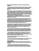
the actin cytoskeleton in response to extracellular signals. FSH is also needed for the formation of lamellipodia and filopodia, with or without the presence of activin A. Activin A determines the cell growth and the massive reorganisation of the actin cytoskeleton, which involves the formation of cortical actin around the plasma membrane of each cell. The cortical actin rearranges to form lamellipodia and filopodia when induced by FSH. This process takes place via the adenylyl cyclase/cAMP pathway, which activates adenylyl cyclase and stimulates cAMP production in granulosa cells and sertoli cells resulting in an increase in intracellular calcium, through voltage-gated and voltage-independent calcium channels. PKA is activated by cAMP, which leads to the phosphorylation of structural proteins, enzymes, and transcription factors causing changes in gene regulation, and therefore resulting in lamellipodia and filopodia formation (Fig. 3).
Fig 3. Model of the signal transductional pathways when FSH binds to the FSH receptor. (Endocrine Reviews 1997).
Changes in the cytoskeletal organisation and cell survival in granulosa cells are also induced when FSH causes activation of the p38 mitogen-activated protein kinase (MAPK) pathway in granulosa cells (Maizels et al, 1998). Phosphorylation of p38 MAPK enzyme, induced by FSH, requires the production of cAMP and activation of PKA. Activation of p38 MAPK is necessary for the phosphorylation of small heat shock protein-27 (sSHP-27), which is induced by FSH, and the rounding of granulosa cells. sHSP are actin capping proteins, which when phosphorylated by FSH, release free actin filaments from the ‘barbed end’ of the polymer, allowing filament reorganisation and stabilisation, and therefore enhancing resistance to apoptosis of granulosa cells. In 1996, Maizels and colleagues demonstrated that FSH can also activate the p42-p44 extracellular signal-regulated kinase (ERK) MAPK pathway in granulosa cell and like the p38 MAPK pathway, requires cAMP production and PKA activation. Activation of ERK is necessary for cellular differentiation and regulation of progesterone synthesis in the ovary (Dewi et al, 2002).
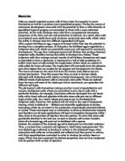
Otsuka and colleagues, in 2000, demonstrated that bone morphogenetic protein-15 (BMP-15), an oocyte growth factor belonging to the TGF-β superfamily, targets granulosa cells to regulate its proliferation and differentiation. It does this by promoting mitotic activity in granulosa cells and in turn stimulating proliferation, inhibiting the expression of the FSH receptor, and stimulating the expression of kit ligand, which is a growth factor present in granulosa cells during follicular development. (Moore et al, 2003). These processes take place via the activation of the Smad1/5/8 pathway. However, expressed in
granulosa cells are follistatin, which inhibits the biological activity of BMP-15 (Otsuka et al, 2001). Thus, BMP-15 regulates ovarian folliculogenesis and follicular function. BMP-6, -7, and -15 have also been found to stimulate FSH secretion and FSHβ subunit transcription in LβT2 cells (Otsuka et al 2002) (Fig. 4).
Fig 4. Effect of BMPs and Activin A on gonadotropin synthesis/secretion in rat primary pituitary cells. (Endocrinology 1996).
IGF-1, IGF-1r (IGF-1 receptors), and FSH receptors, found in granulosa cells of small growing follicles and preovulatory follicles, play critical roles in the development of the follicles. FSH receptors or the FSH-β subunits are important for follicular growth as demonstrated by Burns and colleagues (2001), in which the absence of these components in mice resulted in the impairment of follicular growth beyond the preantral stage. It was shown that in the absence of IGF-1 in mice, severe growth retardation, together with an abnormal follicular growth in the ovary occurred. It is suggested that IGF-1 aids FSH by enhancing the action of FSH in granulosa cells (Orly 2000). However, the exact mechanism by which IGF-1 interacts with FSH is not yet fully understood.
It is suggested that IGF-1 stimulates the phosphoinositide-3 kinase (PI3-K) signalling pathway (Vanhaesebroeck and Alees, 2000), resulting in the activation of phosphoinositide-dependent kinases (PDK) -1 and -2, which subsequently leads to protein kinase B (PKB) phosphorylation and activation. In 2003, Zeleznik et al, determined that the presence of protein kinase B is essential in the stimulation of granulosa cell differentiation by FSH, which acts via the Gsα signalling pathway to induce the expression of aromatase and the LH receptor. However, it was also found that PKB does not have any effect on the proliferation of granulosa cells.
REGULATION OF FSH DURING FOLLICULAR DEVELOPMENT
The regulatory mechanisms that control FSH synthesis, secretion, and functions have not yet been completely understood, but have been under extensive investigation for many years. Regulation of gonadotropin expression involves stimulation by hypothalamic factors (GnRH), inhibitory gonadal feedback (sex steroids), and autocrine/paracrine modulation by intrapituitary factors (activin, inhibin, and
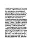
follistatin). The hypothalamus-pituitary-gonadal axis controls the biosynthesis and secretion of LH and FSH, via inhibitory and stimulatory mechanisms (Fig.5).
Fig 5. Schematic model of hypothalamus-pituitary-gonadal axis and factors that regulate gonadotropin biosynthesis and secretion. (Reproductive Endocrinology)
Hypothalamic Control
GnRH secreted by the hypothalamus acts as the main control for gonadotropin expression. GnRH is a decapeptide and is encoded by a gene, which is located on the short arm of chromosome 8. Initially, it was thought that GnRH only stimulated the expression of LH, but now it is assumed that GnRH stimulates the release of both LH and FSH. The anterior pituitary in a pulsatile fashion secretes GnRH, stimulating an increase in the expression of FSH and LH. Many studies have been conducted to confirm the need of a pulsatile release of GnRH for the control of gonadotropin secretion. Use of exogenous pulsatile GnRH treatment restored the secretion of gonadotropin in ewes who were GnRH-deficient (Padmanabhan et al, 2003). Continuous GnRH stimulation leads to a negative feedback effect, which suppresses the secretion of FSH and LH, and their function. This is because pulsatile GnRH causes an up-regulation of GnRH receptors (GnRH-R), while continuous GnRH results in the decline in number of GnRH-R and their sensitivity (Liu et al, 2003). The levels of GnRH in the serum, is difficult to measure in humans, due to the short half-life of GnRH, which is only 2-4 minutes. Studies by Padmanabhan and colleagues (2003 and
Fig 6. Relationship of pulse concordance between FSH and GnRH, and LH (striped boxes). (Endocrinology 1997)
1997) show that pulses of GnRH correlate with LH pulses with 100% concordance, such that there are no LH pulses in the absence of GnRH pulse. Measures of LH pulses indicate the secretion pattern of GnRH, as they both have short half-lives, whereas FSH has a long circulating half-
life. The correlation of FSH and GnRH is also significant with a concordance of 93% (Fig. 6), as each GnRH pulse is associated with an FSH pulse, but there are presence of additional FSH pulses that do not correlate with the pulses of GnRH or LH. These pulses may be due to a separate FSH-releasing factor, but none have been identified yet (Padmanabhan et al, 2003). This suggests that there are two modes of secretion of FSH, a basal and an episodic mode, which are regulated by a dual mechanism (Padmanabhan et al, 1997). The basal mode is the dominant secretion of FSH, while GnRH causes the pulsatile release of FSH in the episodic mode of secretion.
The pattern of secretion of GnRH suggests the presence of a ‘pulse generator’ within the hypothalamus (O’Byrne et al, 1993). The initiator of this pulsatile pattern is yet to be proven, as to whether it is due to GnRH neurones or local electrochemical communications. There are variable frequencies of GnRH pulses in the normal female reproductive cycle. It is estimated that in the early follicular phase, GnRH pulses arise every 94 minutes, which then increases in the late follicular phase to every 71 minutes. In the late luteal phase, GnRH pulses occur at the lowest frequency, of every 216 minutes. In relation to gonadotropin secretion, slow pulse frequencies of GnRH lead to an increase in FSH secretion, and a rapid pulse frequency, causes an increase in LH secretion (Bédécarrats et al, 2003). This suggests that GnRH pulse frequency variations are important in not only the levels of LH and FSH, but also the ratio of LH: FSH secretion. Variations in the pulses of GnRH may be influenced by gonadal steroid production. It was observed in ewes, that the GnRH pulse frequency increased with the production of estradiol, which in turn leads to an increased LH secretion (Molter-Gérard et al, 1999). During the luteal phase, the increased production of progesterone causes a decrease in the GnRH pulse frequency, which results in the secretion of FSH, necessary in the menstrual cycle during the late luteal phase.
Gonadal Steroid Feedback
Two gonadal feedback systems; gonadal steroid and activin-inhibin-follistatin, are involved in the control of LH and FSH biosynthesis and secretion. The mechanism of these feedback systems have an overall inhibitory effect on the gonadotropin serum levels and correlate with the GnRH pulsatility. Oestrogen, progesterone, and androgen are steroids of the gonads and they act on both the hypothalamus and the anterior pituitary, via their receptors, which are found in the gonadotrope cells of the anterior pituitary and within the hypothalamus. The GnRH-containing neurons of the arcuate nucleus in the hypothalamus do not seem to express any steroid receptors and so it has been suggested that the gonadal steroids act indirectly on GnRH release through the neural system (Shupnik 1996). However, GnRH controls the production and release of these steroids via the release of LH and FSH.
Oestrogen causes both inhibitory and stimulatory effects on gonadotropin gene expression. Ovariectomy results in the loss of the oestrogen feedback system, which leads to an increase of circulating LH and FSH and α-subunit, LHβ, and FSHβ mRNA levels. This effect is reversible by the use of estradiol, which acts by decreasing the transcription rate of gonadotropin subunit mRNAs. The inhibitory effects of oestrogen act mainly via the pituitary gland, such that, when an hourly infusion of GnRH was given to rats, in response to estradiol, the secretion of LH was reduced. The inhibitory effects of oestrogen may also act on the hypothalamus pulse generator, such that the GnRH pulsatility is decreased when estradiol is added in rats, leading to a reduction in the frequency of LH pulses. During the LH surge, the stimulatory effect of oestrogen causes an increase in the levels of FSH and LH. In the pituitary gland of the rats, the treatment of estradiol induced the menstrual cycle and the FSH/LH surge. At the level of the hypothalamus, the positive feedback effect of oestrogen causes an increase in GnRH pulsatility with LH surge.
The effect of progesterone has not yet been fully defined and is believed to be dependent on oestrogen priming. Treatment of progesterone, in the presence of oestrogen, in a rat, resulted in increased FSHβ mRNA levels (Attardi and Fitzgerald, 1990), but the treatment of progesterone alone exerted no effects.
Like oestrogen, androgens produce both stimulatory and inhibitory effects on the gonadotropin subunit genes, at the level of the pituitary and hypothalamus, respectively. Experiments carried out in the pituitary of both sexes, in vitro and in vivo, suggest that androgens cause a stimulation of FSHβ mRNA levels and FSH secretion, but have very little effect on α or LH-β gene expression.
Activin-Inhibin-Follistatin System
Activin, inhibin, and follistatin are 3 gonadal polypeptide factors found in the follicular fluid, and each has a selective effect on the FSH gene expression. The mRNAs encoding these peptides have been detected in the pituitary, ovary, testes, and placenta, as well as small concentrations in the brain, liver, kidney, adrenal, and bone marrow. In 1993, Miyanaga et al, suggested that activin, inhibin, and follistatin are produced by the granulosa cells and their production is regulated by the FSH-PKA signalling pathway. Inhibin and activin are closely related, such that, inhibin causes a decrease in the function of FSH and LH, while activin stimulates their functions (Shupnik, 1996). However follistatin leads to a decrease in expression of the FSH-β gene and is less potent than inhibin. These peptides cause changes in LH expression, but mainly effect the expression of FSH.
Inhibin, believed to be the most important of three peptides in the feedback regulation of gonadotropin gene expression (Welt et al, 2003), consists of a α-subunit and β-subunit, which are linked by a disulfide bridge. There are two homologous β-subunits, which bind to the α-subunit to form either inhibin
A (αβA) or inhibin B (αβB). Inhibin specifically inhibits the synthesis and secretion of FSH, which has been demonstrated by many experiments, carried out in vitro and in vivo. However, unlike inhibin, activin does not have a α-subunit, but consists of the two β-subunits, to form a heterodimer activin A (βAβ), activin AB (βAβB), or activin B (βBβB). Follistatin is a polypeptide, which is highly glycosylated and consists of three homologous domains. The secretion of follistatin is regulated by GnRH, in which continuous GnRH stimulates follistatin release, while suppressing FSHβ mRNA (Besecke et al, 1996) (Fig. 7).
Fig 7. FSHβ and follistatin response to pulsatile and continuous GnRH. (Endocrinology 1996).
There are two major types of activin receptors: type I (Act-RI) and type II (Act-RII), both containing many isoforms, which possess protein serine/threonine kinase activity. These transmembrane receptors are located in the cell surface of pituitary gonadotropes. Activin directly binds to the Act-RII, which increases association with Act-RI, to form a complex. This complex formation causes the phosphorylation of Act-RI, which leads to the activation of the Smad transcription factor family, which includes Smad-2, -3, and -4 proteins. Smad-2 and Smad-3, when phosphorylated, form a complex with Smad-4, which translocates into the nucleus to regulate FSH and LH gene transcription, with the help of other co-activators (Dupont et al, 2003). Activin receptors may also activate many other signalling pathways to control the synthesis and secretion of FSH. Act-RI and Act-RII receptor mRNAs have been detected in pituitaries and ovaries of many species including mice, but studies have not yet identified an inhibin-specific receptor or a follistatin receptor. It is suggested that inhibin competes with activin for the activin receptor sites, as inhibin can bind to Act-RII receptors and that follistatin may act through the action of activin or inhibin. Thus, Kogawa et al (1991) suggested that activin induces FSH secretion in the pituitary, which is regulated by the activin binding protein/follistatin. Follistatin binds to the common activin/inhibin β-subunit and so prevents the interaction of activin A with the Act-RII receptor. Therefore, the regulation of FSH by follistatin is indirect and activin-dependent (Bauer-Dantoin et al, 1996). The stimulation of FSHβ mRNA by GnRH is also activin-dependent and blocked by follistatin (Besecke et al, 1996).
When female rats were treated with exogenous estradiol after ovariectomy, the expression of inhibin subunit was suppressed, but with further exposure to estradiol, there was an increase in follistatin gene expression. Direct regulation of gene expression involves GnRH and gonadal steroids, but they can also modulate intrapituitary levels of inhibin, activin, and follistatin, which in turn regulate FSH expression. Besecke and colleagues in 1996 demonstrated that follistatin mRNA and protein expression can be increased by GnRH. The activin-inhibin-follistatin system regulates the biosynthesis and secretion of gonadotropins has been proven by many experiments done in vitro and in vivo model systems. However, a study conducted by Besecke et al (1996), suggests that the presence of an activin/follistatin regulatory loop in the pituitary, which regulates FSHβ mRNA, is responsive to the frequency of GnRH, and regulates synthesis and secretion of FSH in vivo (Fig. 8).
Fig 8. Activin/follistatin regulatory loop for the control of FSHβ gene expression by pulsatile and continuous GnRH release. (Endocrinology 1996).
Bédécarrats et al (2003) have suggested that Müllerian inhibiting substance (MIS) may regulate gonadotropin gene expression at the levels of the hypothalamus, pituitary, and gonads. MIS is a glycoprotein hormone, which belongs to the TGF-β superfamily. Its main action is during the embryonic development, in which expression of MIS causes the regression of the Müllerian ducts, resulting in the male internal reproductive organs. Bédécarrats demonstrated that the addition of MIS to LβT2, a murine gonadotrope-derived cell line, induces an increase in FSH-β subunit mRNA levels.
CONCLUSION
It is now clear that FSH is essential for the proliferation and differentiation of granulosa cells, during follicular development. FSH binds to the FSH-receptor and induces the many FSH-responsive genes, which are mainly mediated through the cAMP pathway, but may also involve the p38 MAPK, ERK, and Smad 1/5/8 pathways. GnRH control of FSH action underlines the importance of the pulsatile release of the hormone from the hypothalamus, which is generated by the ‘pulse generator’. However activin, inhibin, and follistatin also play an important role in regulation of the biosynthesis and secretion of FSH.
Future studies should involve the identification of more FSH-responsive genes, to further enhance our understanding of the function of FSH and its role in, not just follicular development, but throughout the female reproductive cycle. The regulation of FSH requires further investigations, which should be conducted more in vivo.
Fig 9. Schematic diagram of the neuroendocrine regulation system that regulates FSH biosynthesis and secretion. (Archives of Medical Research 2001)
REFERENCE LIST
Front page image:
A ribbon diagram of human FSH with the alpha chain in red and the beta chain in green.
Book:
Voorhis BJV. 1998. Follicular Development. In: Encyclopedia of Reproduction, Knobil E. and Neill JD. Pp 376-383. Academic Press. USA.
Journals:
Attardi, B. and Fitzgerald, T. 1990. Effects of progesterone on the estradiol-induced follicle-stimulating hormone (FSH) surge and FSH beta messenger ribonucleic acid in the rat.Endocrinology 126, 2281-2287.
Bauer-Dantoin AC, Weiss J, and Jameson JL. 1996. Gonadotropin-releasing hormone regulation of pituitary follistatin gene expression during the primary follicle-stimulating hormone surge. Endocrinology. 137: 1634-1639
Bédécarrats GY, O'Neill FH, Norwitz ER, Kaiser UB, and Teixeira J. 2003.
Regulation of gonadotropin gene expression by Müllerian inhibiting substance. PNAS. 100: 9348-9353
Bedecarrats GY and Kaiser UB. 2003. Differential regulation of gonadotropin subunit gene promoter activity by pulsatile gonadotropin-releasing hormone (GnRH) in perifused LbetaT2 cells: Role of GnRH receptor concentration. Endocrinology. Vol. 144(5): 1802-1811.
Besecke LM, Guendner MJ, Schneyer AL, Bauer-Dantoin AC, Jameson JL, and Weiss J. Gonadotropin-releasing hormone regulates follicle-stimulating hormone- beta gene expression through an activin/follistatin autocrine or paracrine loop. Endocrinology. 137: 3667-3673
Burns KH, Yan C, Kumar TR, Matzuk MM. 2001. Analysis of ovarian gene expression in
follicle-stimulating hormone _ knockout mice. Endocrinology 142:2742–2751
Chemyong KO, Grieshaber NA, Inhae JI, and Tae HJI. 2003. Follicle-Stimulating Hormone Suppresses Cytosolic 3,5,3’-Triiodothyronine-Binding Protein Messenger Ribonucleic Acid Expression in Rat Granulosa Cells. Endocrinology. 144:2360-2367
Das S, Maizels ET, DeManno D, St.Clair E, Adam SA, and Hunzicker-Dunn M. 1996. A Stimulatory Role of Cyclic Adenosine 3’,5’-Monophosphate in Follicle-Stimulating Hormone-Activated Mitogen-Activated Protein Kinase Signalling Pathway in Rat Ovarian Granulosa Cells. Endocrinology. 137: 967-974
Dewi DA, Abayasekara DRE, Wheeler-Jones CPD. 2002. Requirement for ERK1/2 activation in the regulation of progesterone production in human granulosa-lutein cells is stimulus specific. Endocrinology 143(3): (877-888).
Dupont J, McNeilly J, Vaiman A, Canepa S, Combarnous Y, Taragnat C. 2003 Activin signaling pathways in ovine pituitary and LbetaT2 gonadotrope cells. Biology of Reproduction 68(5):1877-1887
Grieshaber NA, Boitano S, Inhae JI, Mather JP, and Tae HJI. 2000. Differentiation of Granulosa Cell Line: Follicle-Stimulating Hormone Induces Formation of Lamellipodia and Filopodia via the Adenylyl Cyclase/Cyclic Adenosine Monophosphate Signal. Endocrinology. 141:3461-3470
Grieshaber NA, Chemyong KO, Greishaber SS, Inhae JI, and Tae HJI. 2003. Follicle-Stimulating Hormone-Responsive Cytoskeletal Genes in Rat Granulosa Cells: Class I β-Tubulin, Tropomyosin-4, and Kinesin Heavy Chain. Endocrinology. 144: 29-39
Hsueh AJW, McGee EA, Hayashi M, and Hsu SY. 2000. Hormonal regulation of early follicle development in the rat ovary. Molecular and Cellular Endocrinology. 95-100
Kogawa K, Nakamura T, Sugino K, Takio K, Titani K, and Sugino H. 1991. Activin-binding protein is present in pituitary. Endocrinology. 128: 1434-1440
Liu F, Austin DA, and Webster NJG. 2003. Gonadotropin-releasing hormone-desensitized LbetaT2 gonadotrope cells are refractory to acute protein kinase C, cyclic AMP, and calcium-dependent signalling. Endocrinology. Vol. 144(10): 4354-4365.
Maizels ET, Cottom J, Jones JCR, and Hunzicker-Dunn M. 1998. Follicle-Stimulating Hormone (FSH) Activates the p38 Mitogen-activated Protein Kinase Pathway, Inducing Small Heat Shock Protein Phosphorylation and Cell Rounding in Immature Rat Ovarian Granulosa Cells. Endocrinology. 139(7): 3353-3356
Méduri G, Charnaux N, Driancourt M, Combettes L, Granet P, Vannier B, Loosefelt H, and Milgrom E. 2002. Follicle-Simulating Hormone Receptors in Oocytes? Journal of Clinical Endocrinology and Metabolism. 87: 2266-2276
Miyanaga K, Erickson GF, Depaolo LV, Ling N, and Shimasaki S. 1993. .Differential Control of Activin, Inhibin and Follistatin Proteins in Cultured Rat Granulosa Cells. Biochemical and Biophysical Research Communications. 253-258
Molter-Gérard C, Fontaine F, Guérin S, and Taragnat C. 1999. Differential Regulation of the Gonadotropin Storage Pattern by Gonadotropin-Releasing Hormone Pulse Frequency in the Ewe. Biology of Reproduction. Vol. 60(5): 1224 – 1230.
Moore RK, Otsuka F, and Shimasaki S. 2003. Molecular Basis of Bone Morphogenetic Protein-15 Signalling in Granulosa Cells. Journal of Biological Chemistry. 278: 304 - 310.
O'Byrne KT, Chen M-D, Nishihara M, Williams CL, Thalabard J-C, Hotchkiss J, and Knobil E. 1993. Ovarian control of gonadotropin hormone-releasing hormone pulse generator activity in the rhesus monkey: Duration of the associated hypothalamic signal. Neuroendocrinology. Vol. 57(4):588-592.
Orly J. 2000. Molecular events defining follicular developments and steroidogenesis in the ovary. In
Shupnik MA, ed. Gene Engineering in Endocrinology. Totowa, NJ: Humana Press; 239–275
Otsuka F, Yao Z, Lee TH, Yamamoto S, Erickson GF, Shimasaki S. 2000. Bone Morphogenetic Protein-15. Identification of Target Cells and Biological Functions. Journal of Biological Chemistry. 275: 39523-39528
Otsuka F, Moore RK, Iemura S, Ueno N, and Shimasaki S. 2002. Follistatin Inhibits the Function of the Oocyte-Derived Factor BMP-15*1. Biochemical and Biophysical Research Communications. 961-966.
Otsuka F and Shimasaki S. 2002. A Novel Function of Bone Morphogenetic Protein-15 in the Pituitary: Selective Synthesis and Secretion of FSH by Gonadotropes. Endocrinology. 143: 4938-4941
Padmanabhan V, McFadden K, Mauger DT, Karsch FJ, and Midgley AR Jr. 1997. Neuroendocrine Control of Follicle-Stimulating Hormone (FSH) Secretion. I. Direct Evidence for Separate Episodic and Basal Components of FSH Secretion. Endocrinology. 138: 424-432
Padmanabhan V and Sharma TP. 2001. Neuroendocrine vs. Paracrine Control of Follicle-Stimulating Hormone. Archives of Medical Research. 533-543
Padmanabhan V, Brown MB, Dahl GE, Evans NP, Karsch FJ, Mauger DT, Neill JD, and Van Cleeff J. 2003. Neuroendocrine Control of Follicle-Stimulating Hormone (FSH) Secretion: III. Is There a Gonadotropin-Releasing Hormone-Independent Component of Episodic FSH Secretion in Ovariectomized and Luteal Phase Ewes? Endocrinology. 144: 1380-1392
Richards JS, Russell DL, Ocusner S, Hsieh M, Doyle KH, Falunder AE, Lo YK, and Sharma SC. 2002. Novel Signalling Pathways That Control Ovarian Follicular Development, Ovulation, and Luteinization.
Shupnik MA. 1996. Gonadal hormone feedback on pituitary gonadotropin genes. Trends in Endocrinology and Metabolism. 7: 272-276
Simoni M, Gromoll J, and Nieschlag E. 1997. The Follicle-Stimulating Hormone Receptor: Biochemistry, Molecular Biology, Physiology, and Pathophysiology. Endocrine Revies.18: 739-773
Tilly JL, LaPolt PS, and Hsueh AJ. 1992. Hormonal regulation of follicle-stimulating hormone receptor messenger ribonucleic acid levels in cultured rat granulosa cells. Endocrinology. 130: 1296-1302
Vanhaesebroeck B and Alessi DR. 2000. The PI3K-PDK1 connection: more than just a road to PKB.
Biochem J 346:561–576
Welt CK, Pagan YL, Smith PC, Rado KB, and Hall JE. 2003 Control of Follicle-Stimulating Hormone by Estradiol and the Inhibins: Critical Role of Estradiol at the Hypothalamus during the Luteal-Follicular Transition. Journal of Clinical Endocrinology and Metabolism. 88: 1766-1771
Zeleznik AJ, Saxena D, and Little-Ihrig L. 2003. Protein Kinase B Is Obligatory for Follicle-Stimulating Hormone-Induced Granulosa Cell Differentiation. Endocrinology. 144: 3985-3994