Fundamental human anatomy and physiology.
Fundamental of Atomy and Physiology
Unit 5 Assignment 1
Task 1(P1)
Cell Structure and Function
Introduction
A human body is made up of lots of tiny cells that you can only see under the microscope however this topic will tell you about the individual structure and the function of the cells and the organs within the body.
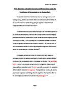
The Eukaryote Cell
Reference:
http://www.bact.wisc.edu/Microtextbook/images/book_4/chapter_2/2-70.gif
Viewed on 25/ 2 / 2008
Cell Membrane
Every cell in the human body is covered by a cell membrane this is a double layer of phospholipids molecules therefore the protein within the cell membrane provides structural support to form a channel for the passage of the materials that acts as a receptor site and functions as a carrier molecule to provide identification maker.
The function of the cell membrane is to separates the materials outside from the inside the cell this is to produce an integrity of the cell and to control passage of the materials in and out of the cell itself.
Reference:
http://training.seer.cancer.gov/module_anatomy/unit2_1_cell_functions_1.html#
Viewed on 25/ 2/ 2008
Nucleus and Nucleolus
The nucleus controls the centre of the cell there fore the thread of the chromatin in the nucleus contains DNA (deoxyribonucleic acid) which makes the genetic materials of the cell.
The nucleolus is a solid area of RNA (ribonucleic acid) which is stored in the nucleus and is the spot of the ribosome structure therefore the nucleus decide on how the cell will function as well as the basic structure of that cell.
Reference:
http://training.seer.cancer.gov/module_anatomy/unit2_1_cell_functions_1.html#
Viewed on 25/ 2/ 2008
Cytoplasm
Cytoplasm is a gel like fluid inside the cell, this is average for chemical reaction this provides a platform so then the other organelles can function properly within the cells.
The cell carries out all the functions of expansion, growth, and replication within the cytoplasm therefore the materials within the cytoplasm moves by diffusion this is a physical process that can only work for a short distance and period of time.
Reference:
http://training.seer.cancer.gov/module_anatomy/unit2_1_cell_functions_1.html#
Viewed on 25/ 2/ 2008
Cytoplasmic Organelles
Cytoplasmic organelles are little organs that are suspended within the cytoplasm of the cell therefore each type of organelle has to define a structure of a specific role in the function of the cell an example of this would be mitochondrion, ribosome, endoplasmic reticulum, Golgi apparatus, Lysosomes,
Reference:
http://training.seer.cancer.gov/module_anatomy/unit2_1_cell_functions_1.html#
Viewed on 25/ 2/ 2008
Mitochondria
Mitochondria is the second largest Organelle, with a single genetic structure which has a double layer of the outer membrane with inner fold known as a cristae this then controls the level of the water and the other materials within the cell therefore the Mitochondria gets rid of (disposes) protein, fat, and carbohydrates and forms a urea.
Reference:
http://www.ivy-rose.co.uk/References/glossary_entry141.htm
Viewed on 25/ 2 / 08
Lysosomes
Is a small body within the cell, the primary function is to digest worn out cell parts and other debris, Lysosomes contain power digestive so then the enzymes can break down into pathogenic such as bacteria therefore the Lysosomes are made up as Golgi complex.
Reference:
http://www.ivy-rose.co.uk/References/glossary_entry128.htm
Viewed on 25 / 2 / 2008
Golgi Apparatus
The Golgi apparatus or Golgi complex is found in most cells this is another packaging organelle like the endoplasmic reticulum, the Golgi apparatus gathers simple molecules and combines (mix) them to make molecules so then it can be packaged in the vesicles so then later on it can send messages out to the cells.
Reference:
http://www.ivy-rose.co.uk/References/glossary_entry90.htm
Viewed on 25/ 2 / 2008
Endoplasmic Reticulum
The endoplasmic reticulum is smooth and rough. The smooth endoplasmic indicates that there are no ribosome's that are attached to the surface of the endoplasmic reticulum and the rough indicates that they are ribosome's which are attached to the surface therefore the smooth and rough endoplasmic reticulum is where the protein and the liquid are produced and concerned about the transport within the cell.
Reference:
http://www.ivy-rose.co.uk/References/glossary_entry204.htm
http://www.ivy-rose.co.uk/References/glossary_entry191.htm
Viewed on 25/ 2/ 2008
Task 2 (P2)
Body Tissues
Tissues is a group of cells that has a similar structure and functions together as a unit for example the intercellular matrix is a non living material which fills the space in between each cells, this may be abundant in some tissues and minimal in other tissues.
Intercellular matrix may contain special substances such as salt and fibres that are unique to a specific tissue and may give those tissues a
The intercellular matrix may contain special substances such as salts and fibres that are unique to a specific tissue and gives that tissue a typical characteristic.
There are four main types of tissue within the body:
* Epithelial
* Connective
* Muscle
* Nervous
Each of these is designed for specific functions.
Reference:
http://training.seer.cancer.gov/module_anatomy/unit2_2_body_tissues.html
Class notes hand out 26 / 2 /2008
Viewed on 26 / 2 / 08
Epithelial Tissue
Epithelial tissue covers and lines the body and its parts, epithelial tissue occurs as a sheet of closely packed cells which covers the surface and lines inside the organs and cavities, this is a barrier that is protective against invasion injury, this is also for incretions or abortions of ...
This is a preview of the whole essay
* Epithelial
* Connective
* Muscle
* Nervous
Each of these is designed for specific functions.
Reference:
http://training.seer.cancer.gov/module_anatomy/unit2_2_body_tissues.html
Class notes hand out 26 / 2 /2008
Viewed on 26 / 2 / 08
Epithelial Tissue
Epithelial tissue covers and lines the body and its parts, epithelial tissue occurs as a sheet of closely packed cells which covers the surface and lines inside the organs and cavities, this is a barrier that is protective against invasion injury, this is also for incretions or abortions of chemical substances for example epidermis, stomach lining.
The structure of each type of epithelium fits its functions i.e. some tissues has a couple of tiny hairs on them called fusilier this is a cell that produces nucleus called a goblet cell.
There are two types of epithelium tissue,
* Simple squamous has one layer of cells.
* Stratified squamous has lots of layers of cells which may be called compound cell.
Reference:
http://training.seer.cancer.gov/module_anatomy/unit2_2_body_tissues1_epithelial.html
Class notes hand out 26 / 2 / 08
Web sight viewed on 26/ 2 / 2008
Connective Tissues
Matrix + fibres = collagen
This makes things strong and stretch.
Connective tissues binds structure together to form a framework and to support the organs and a body as a whole which stores fat, transport substances, protects against diseases and helps repair tissue damage, this occurs threw out the body.
Connective tissue is characterised by an abundant of intercellular matrix with couple of reasonably cells.
Connective tissue are just about able to reproduce but not as fast as epithelial cells therefore most connective tissues has a good blood supply but some don't.
Lots of cell types are found in connective tissue, the three of the most common are the fibroblast, macrophage, and mast cell.
Connective tissue includes loose connective tissue, adipose tissue, dense fibrous connective tissue, elastic connective tissue, cartilage, osseous tissue (bone), and blood.
Reference:
http://training.seer.cancer.gov/module_anatomy/unit2_2_body_tissues2_connective.html
Class notes hand out 26/ 2 / 2008
Web sight viewed on 26 / 2 / 2008
Muscle Tissue
There three types of muscle tissues,
* Skeletal muscle also known as striated muscle this is responsible for voluntary body movements. Muscles will contract because the nerve impulses which are controlled by our conscious threw our central nervous system.
* Smooth muscle also known as non - striated involuntary or plain muscle
These contains protein filaments that are a spindle or cigars shaped this has a single central nuclear dovetails with each other, this then forms sheets which still requires nervous stimulus so then it can effect contraction, this happens threw automatic nervous system.
* Cardiac (hart) muscle moves and pumps the blood around the body. Cardiac muscle only exists within the heart this is an involuntary muscle tissue but its fibre are striated and each cell one nucleus so within its structure it resembles voluntary muscles therefore each cell or fibre has a nucleus.
Muscle tissues are cells that have a special ability to shorten or contract in order to produce movement of the body parts. Therefore the tissue is supplied with the blood vessels.
The cells are called muscle fibres, this is because these cells are longer and slender; these cells are normally arranged in bundles or layers that are surrounded by connective tissues.
Reference:
http://training.seer.cancer.gov/module_anatomy/unit2_2_body_tissues3_muscle.html
Class notes hand out 26 / 2 / 2008
Web sight viewed on 26 / 2 / 2008
Nervous Tissue
Nervous tissue are found in the brain, nerve and the spinal cord and nerve this is so the nervous tissue can coordinate and control many activities within the body an example of this would be when somebody burns themselves they would say ouch, this creates awareness of the environment and plays a major role with the emotions, memory and reasoning but to do all these activities the cell within the nervous tissues needs to be able to communicate with each other by way of sending signals to the nervous impulses.
Reference:
http://training.seer.cancer.gov/module_anatomy/unit2_2_body_tissues4_nervous.html
Class notes hand out 26 / 2 / 2008
Web sight viewed on 26 / 2 / 2008
Task 3 (P3)
Body Organs
Cardiovascular System
Cardiovascular (heart) is in between the chest and lungs with the appendix lying to the left of the body.
* Cardiovascular is a major transport the materials to and from cells
* Takes heat around the body and stays the same in temperature regulations.
* Defence of the body
* Water regulation.
Reference:
http://training.seer.cancer.gov/module_anatomy/unit7_2_cardvasc_heart1_structure.html
Class notes hand out 26 / 2 /08
Web sight viewed on 26 / 2 /08
How the Blood Passes through the Heart
When the blood is deoxygenated it enters through the right atrium and passes through the interior vena cava. The atrium then contrasts and pushes the blood into the right ventricle.
The tricuspid valve is there to make sure that the blood flows the correct way therefore when the right ventricle contracts, it forces the blood through the semi - lunar valve along the pulmonary arteries towards the lungs.
The blood becomes oxygenated within the lungs, the blood then returns to the heat within the pulmonary veins; this then enters the left atrium.
When these contracts it pushes the blood into the left ventricle which has a thick wall because when it contracts it generates high pressure forces the blood through the aorta to the head and the body.
Reference:
Class notes hand out 02 / 04 / 2008
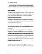
Respiratory System
Respiratory (lungs) each lung lies to one side of the hart filling the chest or thorax.
The lung carries oxygen to the cells and removes carbon dioxide and water from the body.
Reference:
http://training.seer.cancer.gov/module_anatomy/unit9_4_resp_passages.html
Class notes hand out 26 / 2/ 2008
Web sight viewed on 27 / 2 /2008
* The respiratory system consist of the nose, mouth, throat, larynx, trachea, bronchi and lungs, therefore the function of the respiratory system is to help gaseous exchange to take place in the lungs and tissue cells of the body.
* Every cell in our body needs oxygen, therefore the beating of the heart and the movement of our muscles is by cell division however none of these would have been possible without oxygen.
* One breath is enough for oxygen to enter the body but a deeper breath is more affective, therefore the oxygen within one breath will quickly reach the lungs,
* This will then aboard the vehicles that is specially designed for it.
* The blood cells will spread the oxygen threw out the body and it will represent the source of light for each of the cells within the body because the oxygen is getting to the cells.
Reference:
http://www.ivy-rose.co.uk/Topics/Respiratory_System.htm
http://www.brianmac.co.uk/physiolr.htm
Website viewed on 20/5/08
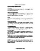
Digestive System
Digestive system includes the stomach, liver, pancreas, duodenum, ileum and colon.
What is a Digestive system?
Digestion is a breakdown and transportation of the solid and liquid food into microscopic substances therefore theses substances are then transported into different areas of the body
The digestive system is a set of organs which transforms whatever we eat into substances that can be used in the body for energy, growth and repair.
Once the food has been broken down by the various chemical processes, and nutrients removed, the rest is excreted as waste.
The whole process involves many different organs and sometimes takes several hours.
There are four stages of digestion:-
* Mouth: ingestion (taking in of food or liquid into the body), chewing and swallowing this is the starch of digestion.
* Stomach: mixing and protein digestion
* Small Intestines: carbohydrates and fat digestion abortion
* Large intestine: waste and excretion
Reference:
http://en.wikipedia.org/wiki/Digestive_system
Website viewed on 28 / 5 / 08
Reference:
http://training.seer.cancer.gov/module_anatomy/unit10_3_dige_regions.html
Class notes hand out 26 / 2 / 2008
Web sight viewed on 27 / 2 2008
Where Does Digestion Start?
The digestion starts within the mouth where the action of the teeth and saliva combined in the first stage of breakdown which is chewing and partially digesting the food so that it can pass threw more easily along the oesophagus therefore the ball of the food that leaves the mouth is known as bolus.
Saliva
* The salvia is a liquid secreted by three pairs of
* The parotid gland placed below the ear
* The submandibular gland and the sublingual gland both of these are placed below the tongue.
* It contains water, mucus and enzyme salivary amylase.
The Three Functions of the Saliva
* To lubricate the food with mucus, making it easier to swallow
* To start digestion: it contains the enzymes salivary amylase, these acts on cooked starch turning it into shorter polysaccharides.
* To keep the mouth and teeth clean.
Reference:
http://en.wikipedia.org/wiki/Digestive_system
Website viewed 29 / 5 / 08
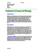
Tongue
The Structure of the Tongue
The tongue is a muscular organ that is covered with a membrane.
It is held in place by attachments to the mandible (lower jaw) and the hyoid bone.
Tiny projections known as papillae cover the top, increasing its surface area and producing a rough texture.
At the side and the base of the tongue, as well as the palate and at the back of the throat there are special areas known as taste buds.
The Function of the Tongue
* The tongue has tree digestive functions: - taste, chewing and swallowing
* Taste: the tongue is covered with thousands of taste buds which are sensitive to salt, sweet, sour and bitter chemicals within the food and drinks.
* They help us enjoy what we eat and drink and also acts as a first line of defense, warning us when food and drinks are foreign matter are off or unfit for human consumption.
* Chewing: the tongue aid is by moving food around the mouth, pushing it between the teeth and covering it with saliva, which contains enzymes that starts the digestion process.
* The food is turned into a partially digested mass known as a bolus.
* Swallowing: when the food is ready to travel to the stomach, the tongue pushes it to the back of the mouth.
Reference:
http://family.innocentdrinks.co.uk/newsletters/2007/august/
http://www.eatatease.com/themouth.html
Website viewed on 30 / 5 / 08
How does food travel from the mouth to the stomach?
* The action of swallowing and through the tube is known as the oesophagus.
* The tongue pushes the bolus to the back of the mouth, towards the pharynx, muscular tube behind the mouth therefore the food passes into the pharynx and down to the oesophagus.
* The epiglottis is a small flap of cartilage which forms part of the larynx (the windpipe) moves upwards and forwards, blocking the entrance to the larynx.
* This stops the food from going down the wrong way and prevents choking.
Oesophagus
The Structure of the Oesophagus
The oesophagus is a muscular tube which leads from the pharynx, at the back of the mouth, to the stomach, the first main organ of the digestion.
The Function of the Oesophagus
* Carry chewed food from the pharynx to the stomach.
* Food moves along it by muscular concentration this is known as the peristalsis.
* The muscle fibers contract and relax which acts like a wave on the tube which pushes the bolus forwards.
* The lining of the oesophagus secretes mucus to ease and lubricate the passage of the food.
Reference:
http://tabacco.blog-city.com/human_digestive_system_overview__anatomy_function_maintenanc.htm
Website viewed on 30 / 5 / 08
Stomach
The Structure of the Stomach
* The structure of the stomach is J shaped, with an elastic organ which expand and contracts depending on what is in it.
* Food enters from the oesophagus therefore the cardiac sphincter is a valve that stops backflow of the stomach contents and leaves though the pyloric sphincter into the duodenum which is the first part of the intestine.
* The wall of the stomach is a combination of layers of muscle fiber with an inner mucus membrane.
* The latter has lots of folds which is called rugae.
* When the stomach if full they stretch out which enabling expansion then they contract when it empties.
The Function of the Stomach
* Digest proteins threw the actions of enzymes.
* Churns food with gastric juices.
* Helps to lubricate the food by producing mucus (from the mucus membrane)
* Absorbs alcohol
* Kills bacteria by producing hydrochloric acid.
Reference:
http://ezinearticles.com/?The-Stomach-and-Digestion&id=613644
http://www.mydr.com.au/default.asp?article=3351
Website viewed on 31 / 5 / 08
Renal System also known as urinary system
Rental system (kidney) the body has one on each side of the posterior wall of the abdomen and the upper poles of the kidney lies just inside the ribs and the left kidney is slightly higher then the right due to the bulk of the liver.
The bladder lies centrally in the lower pelvis at the front.
The function of the kidney is to remove the excess water and salt that eliminates nitrogen- containing waste in the form of urea, the kidney also assists in the production of new red blood cells and involves in the maintenance of the blood pressure.
* Kidney to filter the blood reabsorbs useful materials which are needed by the body and to form urine.
* Ureters to take urine from the kidney to the bladder.
* Urethras to take urine from inside the body (the bladder) to out side. In men the urethras is also a passage to the semen.
* Bladder has an internal sphincter which relaxes and contracts, therefore the opening and emptying the urine into the urethra.
Reference:
http://www.web-books.com/elibrary/medicine/Physiology/Urinary/urinary_system.jpg
Class notes hand out 26 / 2 / 2008
Web sight viewed on 27 / 2 /2008
Nervous System
Nervous (brain) is within the skull of the head.
The function is to receive and interprets information from the environment to control and coordinate the internal organs which is associated to the endocrine system and relaxes the actions which protects the body from injury.
The nervous system informs the brain about what is happening and therefore protects the body; there are two parts of the nervous system: The central nervous system and the peripheral nervous.
The brain is the main unit and it is connected to the rest of the body by the nerve cells which functions as a messenger, carrying information to and instructions from the brain.
They refer back on pain sensation and danger so that the body can respond and remain in what is known as homeostasis this is a stable psychological state.
Reference:
http://www.infovisual.info/03/038_en.html
Class notes handout 26 / 2 / 2008
Web sight viewed on 27 / 2 /2008
Endocrine System
* Endocrine system is a gland that is scattered through out the body.
* The function of the endocrine is to control and coordinate organs therefore maintains the blood glucose, water and salt levels and takes part in reproduction and growth.
* The endocrine system is made up of the endocrine glands that secrete hormones. Although there are eight major endocrine glands scattered throughout the body, they are still considered to be one system because they have similar functions, similar mechanisms of influence, and many important interrelationships.
* Some glands also have non-endocrine regions that have functions other than hormone secretion. For example, the pancreas has a major exocrine portion that secretes digestive enzymes and an endocrine portion that secretes hormones. The ovaries and testes secrete hormones and also produce the ova and sperm. Some organs, such as the stomach
* Intestines, and heart, produce hormones, but their primary function is not hormone secretion. Learn more about endocrine glands and their hormones by selecting one of the following topics.
Reference:
http://training.seer.cancer.gov/module_anatomy/unit6_3_endo_glnds.html
Class notes handout 26 / 2 / 2008
Web sight viewed on 27 / 2 / 2008
Reproductive System
Reproductive system includes ovaries, testis and uterus.
The ovaries are on one of each sides of the posterior wall of the pelvis, below the kidney and the testis are on one of each side of the men's penis in a skin sac called the scrotum outside the body cavities.
The uterus lies centrally in the pelvis with the ovaries on either side connected to the oviduct.
The function of the reproductive system produces gametes which can create a new life when united with a gamete from the opposite sex this assist in growth and is responsible for secondary sexual characteristics.
Female
The organs of the female reproductive system produce and sustain the female sex cells (egg cells or ova), transport these cells to a site where they may be fertilized by sperm, provide a favorable environment for the developing fetus, move the fetus to the outside at the end of the development period, and produce the female sex hormones. The female reproductive system includes the ovaries, Fallopian tubes, uterus, vagina, accessory glands, and external genital organs. Select a topic below to learn more about the female reproductive system
Male
The male reproductive system, like that of the female, consists of those organs whose function is to produce a new individual, i.e., to accomplish reproduction. This system consists of a pair of testes and a network of excretory ducts (epididymis, ductus deferens (vas deferens), and ejaculatory ducts), seminal vesicles, the prostate, the bulb urethral glands, and the penis.
Reference:
http://training.seer.cancer.gov/module_anatomy/unit12_3_repdt_female.html
http://training.seer.cancer.gov/module_anatomy/unit12_2_repdt_male.html
Class notes hand out 26 / 2 /2008
Web sight viewed on 27 / 2 / 2008
Lymphatic System
The lymphatic lies in the tissue spaces between the body cells.
The function of the lymphatic system is to remove excess tissues and proteins from in between the cells and transport the fatty acid from the digestive system, and to defence the body.
Lymph is a fluid similar in composition to blood plasma. It is derived from blood plasma as fluids pass through capillary walls at the arterial end. As the interstitial fluid begins to build up, it is picked up and removed by tiny lymphatic vessels and returned to the blood.
As soon as the interstitial fluid enters the lymph capillaries, it is called lymph. Returning the fluid to the blood prevents edema and helps to maintain normal blood volume and pressure.
Reference:
http://training.seer.cancer.gov/module_anatomy/unit8_2_lymph_compo.html
Class notes handout 26 / 2 / 2008
Web sight viewed on 27 / 2 / 2008
Musculo - Skeleton System
A musclo- skeleton system is a bone of the skeleton and their attached striated muscles from this system.
The function of the musculo skeleton system is effects movement with the nervous system and stores calcium to protect and support vital organs to produce many blood cells.
The skeleton is a hard framework of 206 bones that support and protect the muscles and organs of the human body this is divided into two parts.
The axial skeleton: this supports the head, neck and the trunk this is also known as a torso this consists of skull, the vertebral column, the ribs and the sternum/
The appendicle skeleton supports the appendages or limb and attaches them to the rest of the body it consists of the shoulder girdle, the upper limb the pelvic girdle and the lower limb.
Reference:
http://dspace.mit.edu/html/1721.1/34966/3-051JSpring2004/NR/rdonlyres/91F88A47-AAD8-49A6-897E-AFC44A49EB98/0/chp_biomaterials.jpg
Class notes handout 26 / 2 / 2008
Web sight viewed on 27 / 2 2008
Immune System
The immune system is more scattered then the other system.
The function of the immune system is to defend against invasion by microorganisms, anti cancer role and reject any materials perceived as foreign.
The immune system is also part of our general body defenses against disease. It functions by recognizing viruses and bacteria and converting that information into hormones that activate the immune process.
This response can be both specific, where the body responds only to certain agents and no others as well as nonspecific, where the body works to defend itself any harmful agent that enters the body.
Reference:
http://www.activamune.com/immune_system.PNG
Class notes handouts 26 / 2 / 2008
Web sight viewed on 27 / 2 / 2008
STOMACH WALL