Villi in the small intestine.
Compound epithelia
The principal function of compound epithelia is to protect deeper structures. Multiple layers of cells hamper the passage of materials which is often so characteristic of the simple epithelia. The vagina, mouth, tongue, and the oesophagus are lined by stratified epithelia consisting of layer of squamous, cuboidal or columnar which gradually become flattened by pressure from below as they reach the surface. The lowest layer of cells on the basement membrane actively divides and the older cells are pushed upwards. This type of epithelia is usually a pink colour and often termed mucous membrane. The skin has an outer layer of epithelium similar in structure to the stratified epithelium but with the important addition of a layer of flattened dead cells on the out side. This is known as the epidermis. As the cells advance from the basement membrane they gradually become filled with a protein called keratin and as said to be keratinised or cornified. This layer is vital in keeping micro organisms from invading deeper structures and has a water proofing effect on the skin. Skin can be variously coloured with pigment produced by pigment cells in the lowest layer. A pigment melanin darkens under the influence of the sun. The numbers of pigment cells in the skin is genetically inherited although they are capable of dividing during exposure to sunshine.
Connective tissues
These tissues are the most widely distributed in the body and lie beneath the epithelial tissues, connecting different parts of the internal structure. Various types of cells lie in background materials known as a matrix. The matrix may be liquid as in blood, jelly-like as in areolar tissues, firm as in cartilage or hard as in bones. The matrix of a tissue is usually secreted by the connective tissue cells. The functions of these tissues are to transport materials, give support, strengthen and protect. Many tissues contain different fibers secreted by the cells to provide special characteristics.
Connective tissues are found in:
Blood
Cartilage
Bones
Areolar tissue
Adipose tissue (fatty tissue)
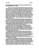
Blood human blood
Blood is a specialized biological fluid consisting of red blood cells (also called RBCs or erythrocytes), white blood cells (also called leukocytes) and platelets (also called thrombocytes) suspended in a complex fluid medium known as blood plasma. Blood plasma is the liquid component of blood, in which the blood cells are suspended. It makes up about 55% of total blood volume.By far the most abundant cells in blood are red blood cells. These contain hemoglobin which gives blood its red color. The iron-containing heme portion of Hemoglobin facilitates hemoglobin-bound transportation of oxygen and carbon dioxide by selectively binding to these respiratory gasses and greatly increasing their solubility in blood. White blood cells help to resist infections, and platelets are important in the clotting of blood.
Blood is circulated around the body through blood vessels by the pumping action of the heart. Blood is pumped from the strong left ventricle of the heart through arteries to peripheral tissues and returns to the right atrium of the heart through veins, blood then enters the right ventricle and is pumped through the pulmonary artery to the lungs and returns to the left atrium through the pulmonary veins, blood then enters the left ventricle to be circulated again. Arterial blood carries oxygen from inhaled air in the lungs to all of the cells of the body, and venous blood carries carbon dioxide, produced as a waste product of metabolism by cells, to the lungs to be exhaled.
Cells
Cartilage
Cartilage is a specialized connective tissue that provides for both strength and flexibility. It is mainly found in the form of hyaline cartilage (hyalos means "glass" in Greek), which is so named because of its smooth, glassy bluish-white appearance when fresh. Cartilage forms the precursor for the vertebrate skeleton in the embryo, which is replaced by bone in the neonate. The exception to this is the articular surfaces of bones (articular surfaces are where two bones move against one another) involved in joints and the ventral ends of the ribs. Hyaline cartilage is also what makes up the cartilage in the nose, bronchi, larynx and trachea. The cells of cartilage are called chondrocytes and are found in spaces (called lacunae) surrounded the extra cellular cartilage matrix. It does not contain blood vessels and is nourished by diffusion from underlying bones.
Bones
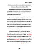
Long bone or spongy bone
Bones are rigid connective organs that make up the skeleton of vertebrates. Bones are primarily comprised of osseous tissue which may also be referred to as bone or bone tissue.
Bone is much harder substance than cartilage but it can be worn away by friction. The ridge matrix has two major components:
calcium salt, which form around collagen fiber and give bone its hardness.
Collagen fiber, which offer some ability to band under strain abd prevent bone from being too brittle and there fore being very likely to fracture.
Osteocytes or bone cells are trapped in the hard matrix in concentric rings called lamellae. A system of these rings is known as haversian system or osteone. Blood vessels and nerves pass through the hollow centre of each osteone. Bone is designed to bear weight and limb bones are hollow, like girders. Bone is also used to protect vital weakers tissue such as the brain, lungs nad heart.
Areolar tissue
Areolar connective tissue
Areolar connective tissue is the most widely distributed connective tissue. It can be found in the skin as well as in places that connect epithelium to other tissues. The areolar tissue is found beneath the dermis layer and is also underneath the epithelial tissue of all the body systems that have external openings. It is also a component of mucus membranes found in the digestive, respiratory, reproductive, and urinary systems. It also surrounds the blood vessels and nerves. Areolar connective tissue holds organs in place and attaches epithelial tissue to other underlying tissues. It also serves as a reservoir of water and salts for surrounding tissues. Almost all cells obtain their nutrients from and release their wastes into areolar connective tissue.
Adipose tissue adipose tissue under the skin
Adipose is a technical term for fat tissue and it is a variation of areolar tissue in which the adipose or fat cells have multiplied to obscure other cells and fibers. When mature, an adipose cell becomes so loaded with fat that the nucleus is pushed to one side and as fat is translucent the cell takes on a distinctive ‘signet rings’ appearance. Adipose tissue is common under the skin and around organs such as heart, kidney and parts of digestive tract. It helps to insulate aganieat cahnges of external temperature, act as a hydraulic shock absorber to protect against injury and is high- energy strong depot.
Muscle tissue
Muscle is an exitable tissue because it is capable of responding to stimuli. There are three different types of muscles in human body:
- Striated
- Non-striated
- Cardiac
Each is composed of muscle fibers capable of shortening or contracting and returning to their orginal state known as relaxation. Contraction causes movement of the skeleton, soft tissue, blood or specific material such as urine, food and faeces. Muscle has both blood and nerve supplies. Muscle activity generates heat and contributes to maintaining the body temperature.
Striated muscle
Striated muscle is also call skeletal muscle.it usually attached to the skeleton although some facial muscles are attached to skin. Skeletal muscles are used to create movement, by applying force to bones and joints; via contraction. They generally contract voluntarily (via somatic nerve stimulation), although they can contract involuntarily through reflexes
Muscle cells (also called fibers) have an elongated, cylindrical shape, and are multinucleated (in vertebrates and flies). The nuclei of these muscles are located in the peripheral aspect of the cell, just under the plasma membrane, which vacates the central part of the muscle fiber
Skeletal muscles have one end (the "origin") attached to a bone closer to the centre of the body's axis and this is often but not always a relatively stationary bone (such as the scapula) and the other end is attached across a joint to another bone further from the body's axis. Contraction of the muscle causes the bones to rotate about the joint and the bones to move relative to one another (such as lifting of the upper arm in the case of the origin and insertion described here).
bone
Non-striated nucleus
Although this type of muscle tissue ( aslo called involuntary, smooth or plain muscle) still contains the protein filaments, they do not lie in an ordered pattern and therefore do not produce the banding characteristic of striated muscle. The muscle fibers are spindle – or cigar shaped with single cental nuclie, and dove tail with each other.this type of muscle tends to form sheets and although still requiring nervous stimulation to effects contraction, thin is not under conscious thought, but supplied by autonomic nervous system. This type of muscle id found around hollow internal organs such as the stomach, intestine, iris of the eyes, bladder and uterus. Non- striated muscle frequently occurs in two sheets running in different diractions. In the digestive tract, one sheet runs circularly around the intestines while another outer sheet runs sown the length. The two sheets are said to work antagonistically to propal the food contents down the tract. This is known as peristalsis. In the iris of the eyes, one set of muscle runs radially like o spokes of a wheel while other outer set runs circularly around the central pupil. This arrangement allows for the control of light entering the eyes and the pupil is said to be dilated or constricted.
Connecting tissue
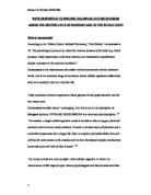
Smooth muscle cells
Cross striation Intercalated discs
Cardiac muscle myocytes
This muscle is found only in four chambers of heart. It is said to be myogenic because it is capable of rhythmically contracting without receiving any nervous stimuli and in this it differ from other muscle. The muscle branch repeatedly to form a network through which contraction spreads rapidly. Each cell has a central nucleus and is both horizontally and vertically striped. The divisions between cells are known as intercalated discs and are specially adapted for transmission of impulses. Under normal healthy circumstance, cardiac muscles is not allowed to contract myogenically because the atrial or upper chamber muscles has a different contraction rate to that of lower ventricular muscles and this would lead to inefficient and uncoordinated heart action. The automatic nervous system controls the rate of contraction in order to adapt the flow of blood to specific circumstances such as rest and exercise.
Branching network
Nervous tissue
Nervous tissue is only found in nervous system and consist of brain, spinal code and nerves. Receiving stimuli from both external and internal sources, it serves to create consistency, co-ordination and communication between different parts of the body. The nervous system interprets stimuli from the senses organ so that vision, hearing, smell etc become apparent.
Nervous is composed of:
Neurones- highly specialised nerve cells which transmit nervous impulses. They are present only in brain and spinal code, but their long process forms the nerves.
Neuroglia- connective tissue cells intermingled with neurones in the brain and spinal code that offers support and protection.
Nucleus
Bibliography
BTEC national health and social care certificate text book
Wikipedia
http://www.uoguelph.ca/zoology/devobio/210labs/ct2.html
A Level biology text book
Michael Kent advanced biology
Oxford ISBN 0-19-914105-9