Apert syndrome.
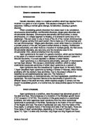
Apert syndrome is a condition, were the person has abnormal growth of the skull and face. This is because of a mutation of the fibroblast growth factor (FGF) receptor 2 (FGFR2). The mutation of this gene results in a craniofacial problems, and what separates Apert syndrome from the other craniofacial syndromes is syndactyly of the limbs. Syndactyly is the fusion of the soft tissues, of the fingers and toes. The level of syndactyly can very, from the fusion of the three digits, to complete fusion of the bones and nails of all five digits. There are three types of Apert synalactylies, depending on the level of fusion; they are types 1-3. Type 1 is were only the index, long finger and ring fingers are fused. Type 2, the fingers are all fused together. Type 3, all the fingers or fused together, and the thumb is covered by skin. In regards to the toes, it’s the soft tissue which is fused, but bone fusion can occur, but is rare. The big toes are usually shorter and curve away from the foot. The foot is also smaller then normal. (see figure 1)
.
Legend: a shows type one Apert syndactyly, were the middles digits or fused together, but the thumb is free, b shows type two Apert syndactyly, were the thumb is fused and all the digits are fused, c shows type three syndactyly, were the digits or fused, and the thumb is covered in skin. Image d shows fused toes of an Apert foot.
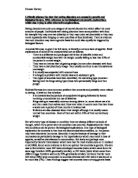
The main feature of Apert syndrome is that there is an abnormal growth of the skull, and face. The skull of a person with Apert is described as Brachycephaly, which is the skull appears short from the front to the back. There is also a lack of development in the midface, described as concaves. This is due to the fact, that when the skull grows, or is enlarged by surgical proceedings the middle third of
the face grows slower. The smaller misfile is known as midfacal hyperplasia. The Brachycephaly structure is the result of the coronal structures been fused, so that the fontral bone is not separated from the bilateral parietal bone. The fantanellous is the soft tissue which separates, them parts of the skull.
Figure two: Apert syndrome.
Legend: A two Children’s faces with Apert syndrome
The skull of normal babies, the top of the skull is the anterior fantanelle; at the back of the head is the posterior fantanelle. The fantanellous allows a temporary change in the head shape, during the birthing process; this makes it easier for the baby to pass through the birthing canal.
The fusion of the growth lines, of the skull means that the bones around the effected area are unable to at the correct rate, causing the Brachycephaly. The abnormal growth of the skull causes the orbits around the eyes to become shallow, and the eyes will appear to bulge. This can cause a problem known as exarbitism were the eyelids are not able to close correctly.
In normal infant’s skulls, three are the fontal bone, occipital bone, two partial bones and two temporal bones. They are known as the cranial bones. In between them bones are cranial sutures, which hold the bones together. The cranial is made of a strong, fibrous, elastic tissue. The cranial bones do not undergo ossification (fusion), until the child is between 12-18 months, producing the adult skull.
The fontanel sutures are flexible, throughout childhood allowing the skull to expand with the rapid growth of the brain. It also prevents constriction and protects the brain from miner impacts.
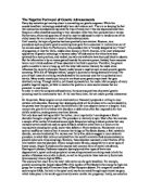
In Apert syndrome the bone has grown together were the fontanels and sutures should be, without the flexibility which the fantanelle and sutures allows, the brain of Apert children become constricted within the cranial bones, and can not grow adequately without surgical procedures. (see figure 3)
Figure 3: An artist’s impression of the skull of a normal Childs skull.
Legend: A Childs skull, and the names of different areas, of the Childs skull.
Figure 3.5: An artist’s impression of a skull of an Apert Childs skull.
Legend: A Childs skull, and the names of the different areas of the Childs skull.
The mutation which results in Apert syndrome is the fibroblast growth factor receptor 2, on chromosome ten. The Mutation occurs on two adjacent residues, the Ser262 is changed to Trp, and Pro253 is changed to Arg, between the linker regions between Ig11 and Ig111 of the genes ligand binding domain. This is referring the fact that that the nuclear hormone receptors are activated by ligands, for the transcription process. This change in the amino acids, changes the primary structure of the amino acid structure, which when folds into the secondary structure, the correct links between the amino acids dose not occur. This effects the ligand binding domain, of the three dimensional shape of the hormone receptors. It is those hormone receptors which are activated by ligands for the transcription process.
Transcription is the process, were massager RNA (mRNA) is produced from DNA. The mRNA contains the genetic information, which is encoded by the enzyme transcriptase.
The process of ligand activated transcription plays an important role, in regulating the gene expression, through the interaction with the DNA sequences, producing a ligand-DNA complex, on a target gene. The ligands play an important role in producing different orientations of the DNA. This influences the property of the receptor, to discriminate and identify different binding molecules. The change of ser and pro, been changed to Trp and Pro, effects the transmembrane receptors, which bind to the FGF ligands, because of the ability of discrimination and identification is effected. Those mutant receptors have an increase affinity for certain FGF ligands, resulting in signalling, even when FGF ligands availability is low. This increased affinity is what causes the fusion of the bones in the skull, and conjoining of the digits.
Conclusion.
Apert syndrome is a rare genetic disorder, which can be the result of either an inherited mutation, or a random mutation on the FGFR2 gene. The mutation is the change of to adjacent amino acids. This change affects the primary structure of the protein, which results in abnormal folding into the secondary and tertiary structure of the transmembrane receptor, which binds to FGF ligand. This change causes the receptor to have an increased affinity for the FGF ligand. The increased affinity stimulates the over growth of the bones in the skull and face, causing them to fuse. There is also fusion in the digits, this is the soft tissue and can sometimes be the bones as well. Those problems are classified as craniosyntosis, mid face hyperplasima and syndactyly.
The conditions such as craniosynostosis require surgical procures to allow the brain to grow. It Important as the skull and mid face abnormally slow.
Reference
Abigal.A.Salyers and Dixie D.Whitt (2001), microbiology: Diversity, Disease, and the environment, Fitzgerald Science press, first edition, Bethesda Maryland.
Michael T. Madigan and John M Martinko(1970), Brock: Biology of Microorganisms, Prentice Hall, Eleventh Edition, Southern Illinois University Carbondale.
Biochemistry (second edition), Donald Voet and Judith A. Voet, edited by Judith Rose. John Wiley and sons Inc.
Molecular Mechanisms and Keinetics between DNA, and DNA binding ligands Biophysical Journal, Jan 2005, Sidchka, Andy, Toensins katia, Rainer, Wilking, Sven David.
Apert syndrome mutations in fibroblast growth factor receptor 2 exhibit increased affinity for FGF ligand, J. Anderson, HD Burns, P Enriquez-Harris, AOM Wilkie and JK Heath, Human Molecular genetics, vol 7, pg 1475-1483, (1998) Oxford University Press.
Clinical assessment and multipecialty, management of Apert syndrome, Lawrence C. Kaplan MD, Clinics in plastic surgery vol 18, no 2, April 1991.
Childrens cronianofacial Association: A guild to understanding Apert syndrome, Jeffrey Fearen, MD, corolyn Johnson M.S Ed, June 1993. (leaflet)
Wilkie, A.O. M ET AL. Apert syndrome results from localized mutions of FGFR2 and it’s allelic with Crouzan syndrome, Nature of genetic, vol 9, 1995, pg 165 -172.
Internatal Journal of development in biology, vol 46, 2002, pg 817-825, Dr. P. Dvorak. Development of molecular Embryology: Targeted disruption of Fibroblast growth factor receptor, blocks maturation of visceral endoderm and cavitations in mouse embryonic bodies.
Prodceding of the National Academy of sciences of United states of America, June 19, 2001, vol.98, no 13, pg 7182 – 7187, Joseph Schlessinger, Yale University School of Medicine, New Haven.
Figure1: www.thecraniofacialcenter.org/.../ apert_hand.jpg
Figure 2
www.stronghealth.com
Figure 3;www.headlines.org.uk/ images/SkullWeb.jpg