-T cells which have not yet met their specific antigens are called naïve t cells.
-naïve CD8 t cells will later develop into cytotoxic t cells if they are activated.
Naïve t cells are released from the thymus and circulate through the blood stream and through lymphoid organs through a process of migration.
As they migrate they become temporarily bound to antigen presenting cells (APCs), usually dendritic cells. This binding is caused by adhesion molecules.
All cells have major histocompatibility (MHC) complexes on their surfaces. Class 1 MCH molecules express a sample portion of the proteins made within the cell, forming a groove around this peptide molecule. Under normal cellular conditions this peptide will be a fragment of the usual cell constituents. If the cell is virally infected, the virus would be using cellular equipment to synthesise its DNA. In this case a foreign peptide strand would be displayed in the groove of the MHC molecule.
Naïve cells sample MHC- peptide complexes on the surface of the APCs. Temporary binding allows the naïve cell to sample many different MHC peptide complexes in a short period of time.
Activation
The naïve t cell is activated when it encounters a foreign peptide bound to an MHC complex. This alerts the naive t cell that the target cell has become virally infected.
Activation causes stabilisation of the transient bonds formed by cell adhesion molecules. Activation requires;
- Simultaneous stimulation from a co-stimulatory signal, produced by the APCs.
- Presence of the CD8 molecule on the naïve t cell which binds to the MCH 1 molecule.
On activation;
- the t cell starts to divide to produce the differentiated cytotoxic t cell
- produces interleukin 2 (IL-2)
The IL-2 stimulates the proliferation of the mature cytotoxic t cell.
Binding
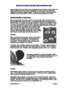
Figure 1. Diagram to show binding of the cytotoxic t cell to the antigen presenting cell.
The activated cytotoxic t cells bind to MHC-peptide complexes using non-specific interactions between LFA-1 and CD2 adhesion molecules. If the cell recognises the antigen this triggers an increase in the affinity of the LFA-I on the t cells for their ligands on the antigen complex. Binding signals a reorientation of the t cells cytoskeleton, to focus lytic granules on to the point of contact with the target cell.
Destruction of the Target Cell
Cytotoxic t cells programme the cell to destroy itself from within. This is called apoptosis.
This is done by releasing the lytic granules (a process which is calcium dependent).
The lytic granules contain 2 classes of cytotoxic substances; Perforins and Granzymes.
Perforins- create pores in cell membrane.
These pores allow water and salts to pass in and out of the cell destroying normal osmotic conditions and so causing cell death.
Granzymes- These enter the cell through pores made by the perforins. Granzymes do not directly cause apoptosis, they trigger of an enzyme cascade. The end product of which is a cleaved inhibitory protein called ICAD. This enzyme is thought to cause DNA degradation and so apoptosis.
Most degradation occurs through the combined action of the above mentioned enzymes however. The cytotoxic t cells can also cause degradation which is calcium independent. I.e. with out the breakdown of lysosomes.
-Fas in target cell membrane binds to Fas ligand on the cytotoxic t cell .This leads to activation of caspases. These cause breakdown of the target cell's protein synthesis mechanisms by cleaving all aspartic acid residues.