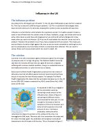
The virus is a globular protein surrounded in a lipid bilayer. Inside the bilayer are 500 molecules of hemagglutinin, 100 molecules of neuraminidase, some 3000 molecules of matrix protein and 8 pieces of RNA. Each of the 8 RNA molecules is associated with many copies of a nucleoprotein and several molecules of three different RNA polymerases.
When the virus enters the body through the respiratory tract of a person, the hemagglutinin molecules bind to carbohydrates on the glycoproteins of the epithelial cells .The virus is then engulfed into the host cell by receptor mediated endocytosis. The RNA of the virus then enters the nucleus of the host cell where fresh copies of the virus are made. These copies then ‘leak’ out into the cytoplasm where some are used as mRNA molecules to translate the proteins of fresh viral particles. Fresh viruses then bud off from the plasma membrane of the cell spreading infection to uninfected cells.
Once someone has been infected the first part of the immune system to take effect is the non-specific immune system. Inflammation occurs because mast cells in tissue beneath the skin detect a foreign body and respond by secreting histamine. This makes the capillaries adjacent to the tissue porous allowing the movement of plasma proteins and phagocytes from the blood to the area of infection. The extra blood supply makes the area look red, while the leakage of fluid from the blood makes the area swollen.
Phagocytosis cannot take place without two different types of white blood cells - neutrophils and macrophages. Neutrophils are granulocytes they mature and are stored in the bone marrow, increasing in number in response to infection, and often only survive for hours or days due to death occurring after engulfing a few micro-organisms by phagocytosis meaning they are continually replaced. However, macrophages mature in the bone marrow, but reside in the major organs. In the liver and are much longer lived.
The process of phagocytosis begins with the gathering of neutrophils at the site of infection by chemotaxis. They are attracted by complement proteins, activated by the invading body, as well as chemicals released by the virus and other substances such as histamine. Receptor proteins on the neutrophil cell surface membrane bind to the surface compounds on the influenza virus. This adherence causes the neutrophil membrane to fold in on itself surrounding the virus in what is known as a phagosome. The binding tends to happen after the virus has been labelled by complement proteins or by antibodies (involving macrophages). Lysosomes situated in the neutrophil cytoplasm then bind to the phagosome forming phagolysosomes, releasing highly toxic free radicals, as well as hydrogen peroxide to kill the virus. The cell wall of the virus is digested by lysozyme, while proteases, and nucleases, along with other enzymes digest the remains of the virus. The products are then mopped up and absorbed into the neutrophil cytoplasm.
The specific immune system works alongside the non-specific immune system described above. During the initial infection some of the virus particles will have been ingested by macrophages. This ingestion, however, does not completely destroy the virus, but instead breaks down the surface molecules of the virus cell membrane into small chunks. These act as antigens and the macrophages display them on their cell surface membrane in grooves within the MHC protein. This is known as antigen presentation. An antigen is any macromolecule, for example, proteins and polysaccharides, to which an immune response (a response to a specific pathogen resulting in the production of a range of cellular and chemical agents of defence directed at that pathogen) may be made. Antigens are divided into self antigens to which a response will not usually be made, and foreign antigens, for instance, viral coat proteins and toxins, to which an immune response will usually be made. On antigenic proteins are specific regions called epitopes which are recognised by one specific antibody.
T4 Helper cells (lymphocytes) then bind onto the viral fragment on the surface of the macrophages. There will only be a few of these T4 cells with the correct receptor proteins to recognize the viral fragment. This process is called clonal selection. Infected cells release cytokines, which cause blood vessels in the infected area to dilate and the infected area to become inflamed. T4 cells release opsonins that label infected cells. These cells will be sought out by killer T cells (T8 cells) and will undergo phagocytosis. The T4 cells also release lymphokines that also cause inflammation. Lymphokines released by the T4 cells initiate the process of clonal expansion. The selected T4 and T8 cells with the receptor cells complementary to the virus proteins are reproduced rapidly by mitosis. The infected cells are now tracked down by the killer T8 lymphocytes. These T8 cells can kill virus-infected cells. Receptor proteins on the T8 cells recognize proteins on the virus-infected cells. Once a cell is found, the T8 cell inserts a perforin protein into the membrane of the cell creating a hole in effect. Then they inject proteases and nucleases into the cell to kill it by digesting its proteins. Lastly they release interferon, which is a viral reproduction inhibitor. This prevents the virus from spreading any further.
The lymphokines released by the T4 lymphocytes also stimulate B-cell activation. B-cells undergo clonal selection until the correct B-cells are found. Once found the B-cells undergo clonal expansion by mitosis. As mitosis produces genetically identical cells when reproduced, each of these B-cells codes for the same proteins and hence the same antibody. The plasma B-cells are the ones that produce antibodies and they produce thousands per second. These antibodies can detect the virus fragments and hence help to stop the infection, usually by either agglutinating the cells for later phagocytosis, or ‘sticking’ to the cells to immobilize them. After the infection, most plasma B-cells die off but a few memory B-cells remain patrolling the blood. In the future, if the body is infected again with the same virus, the memory B-cells will detect the virus and produce a faster, stronger and longer lasting response meaning that you will probably not even feel the symptoms of the flu before the infection is over.
Words: 1297