From Graph I, the point of incipient plasmolysis for the potato tissue sample is 0.26 mol dm-3 and the point of incipient plasmolysis for the swede sample is 0.40 mol dm-3. Using Reference graph II the solute potential of each tissue can be calculated. The solute potential for the potato tissue sample at the point of incipient plasmolysis was 725 kPa. The solute potential for the swede sample at the point of incipient plasmolysis was 1125kPa.
In a plant cell the equation, Water potential = Solute potential + pressure potential however, at the point of incipient plasmolysis Pressure potential = 0, therefore water potential = Solute potential. Therefore, in theory if the potato was placed in a solute solution of 725kPa, and the swede was placed in a solute solution of 1125kPa, there should be no change in mass. This is because there would be no net movement of water as the two solutions solute potentials are the same and equilibrium is already established. The above results show that the potato has a higher water potential then the swede.
The results from the appearance test (Table I) show that a solution with a high water potential will cause the potato and swede chips to become turgid whereas a solution with a low water potential will cause the potato and swede to become flaccid (plasmolysis). From this, we can estimate what the solute potentials of the swede and potato samples are. When the potato chip or swede chip became turgid, its mass increased because water would enter into the cells via osmosis. In contrast, when the tissue was flaccid its mass decreased as water would leave cells and into the solution to reach equilibrium.
When the potato and swede chips were submerged into a hypotonic solution where it has a high water potential (e.g. 0.0 mol dm-3, 0.2 mol dm-3 and 0.4 mol dm-3) the potato and swede chips became very turgid and increased in mass (Table I). This is because there was there was a net movement of water from an area of high water potential (outside the cell) to an area of low water potential (inside the cell).
The potato and swede chips are essentially plants and these cells contain a cell membrane and cell walls. This is an important feature of plant cells as it prevents the cells from bursting when the chip is placed into a hypotonic solution; the protoplast expanded making the cell turgid because the pressure and water potential increased. At this point water potential = solute potential + pressure potential. As a consequence of water moving from the hyposolution into the cells in order to reach equilibrium, the mass of the chip increased as well.
The potato chips and swede chips became flaccid and decreased in mass when submerged in hypertonic solutions with low water potentials (0.6 mol dm-3, 0.8 mol dm-3. 1.0M). This is because in a hypertonic solution, there was a net movement of water from the potato and swede chips cells into the surrounding solution. This caused the chips to become flaccid and decrease in mass. At this point the water potential = solute potential, this is because the protoplast is exerting no pressure on the cell wall and is 0. From these observations, we can deduce that the potato and swede chips have a higher water potential then the test solutions because they decreased in mass.
The solution which caused the chips to gain the most mass and become the most turgid was 0.0 mol dm-3. The potato chips gained an average mass of 19.3%, (0.44g), and the swede chips gained an average mass of 18.6%, (0.38g) when submerged in this solution. The solution which caused the chips to lose the most mass and become the most flaccid was 1.0 mol dm-3. The potato chips lost an average mass of 34.2%, (0.74g) and the swede chips lost an average mass of 23%, (0.45g) when immersed in this solution.
Overall, the solutions which caused an increase in mass for both chips were solutions 0.0 mol dm-3, 0.2 mol dm-3, and 0.4 mol dm-3. The solutions which cause a decrease in mass for both chips were solutions 0.6 mol dm-3, 0.8 mol dm-3, and 1.0 mol dm-3.
From graph I and looking at the line of best fit I can predict that if the sugar solutions were to continue past 1.0 mol dm-3 the line of best fit would eventually level out and become constant. This is because the potato and swede cells would run out of water. So no matter how much more the solute potential of the solution would become, the cells would all become as flaccid and decrease in mass as much as it could, until it couldn’t become any more flaccid or decrease in mass as the cell would have run out of water.
A food test was carried out simultaneously to help explain the differences in results between the swede and potato. From table II the starch test showed that there is more starch present in the potato than the swede. This is because the iodine solution turned the potato sample completely black whilst only parts of the swede changed colour, indicating that more starch is present in the potato. Starch is insoluble therefore it does not affect the water potential in cells. However sugar is soluble and therefore does affect the water potential by lowering it.
The sugar test showed that reducing and non-reducing sugars are present in the potato and swede, the only difference being that the swede contains more of the sugars. This is because the swede sample turned into a yellow/orange colour when the Benedict’s was added, whereas the potato sample only turned into a yellow colour. The Benedict’s turning into a deeper shade indicates that the swede has more reducing and non-reducing sugars present. This is the reason why the potato has a higher water potential than the swede which is why the swede’s line of best fit on graph I is higher than the potato’s line of best fit.
For this experiment care was taken throughout the methods. A labcoat and goggles were worn all times of the experiment for safety, as different substances were being dealt with that might be harmful. Extra care was taken especially when dealing with glass and sharp objects to avoid being cut or breaking of equipment. As well as this, precaution was taken when dealing with the reagents e.g. hydrochloric acid as it is an irritant to the skin. Water was heated to 80oC, so caution was definitely taken when placing and removing test tubes from the water as one can be burned easily. Unneeded materials such as the potato remainders and solutions were discarded properly. Reagents were put away correctly, and the used equipment was washed. By doing so, the experiment was carried out safely and efficiently producing fair results.
Evaluation
I carried out this investigation accurately and fairly. To minimise errors from occurring, I made sure I used the correct equipment and followed the protocols exactly. The safety procedures there also followed so there were no accidents during the experiments. The results I obtained are fairly reliable having very little variation between samples I and II and their changes in mass. However, there were also several anomalies in my data sets caused by large differences between samples I and II and their changes in mass. These anomalies are circled on graph I.
From table V, the most unreliable pair of results for the potato sample was at 0.8 mol dm-3, where the difference in the changes in mass was 10.6% (0.28g). These are the most unreliable pair of results as they have the largest difference between the changes in mass for potato samples I and II. For the swede sample, the anomalous result was at 0.0 mol dm-3 (distilled water), where the difference in the changes in mass was 11.8%, 0.19g. Refer to table V.
Despite the presence of these anomalous results, there were also reliable results which can be observed where the differences between the percentage change in the mass of the tissues was very small. The most reliable pair of results for the potato was at 0.4 mol dm-3, the difference between the changes in mass was 1.67%, 0.04g. For the swede, at 0.6 mol dm-3, the difference between the changes in mass was 0.09% (0.01g).
On graph I, the line of best fit drawn is subjective. If drawn inaccurately, the point of incipient plasmolysis may not be correct. However, a firm trend can still be observed on the graph as the line of best fit was drawn through the reliable results and missing the anomalous results with a large the large deviations. Therefore, the line of best fit drawn on the graph is fairly accurate.
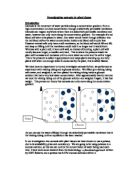
In this experiment there were many sources for possible errors that could have affected the reliability of the results. These errors were most likely to be caused by many factors associated the experimental methods and the way it was carried out. The most prominent error was that when the chips were placed inside the boiling tubes, they were stuck together or onto the side of the boiling tube. This probably occurred in solutions which produced anomalous results. If either of the situations occurred, this would affect osmosis as the surface area of the chips is reduced. The larger the surface area the faster osmosis can occur as the water molecules can cross it at any moment. Therefore, if the surface area of the chips is reduced then this decreases the rate of osmosis. If the rate of osmosis is decreased then fewer water molecules move in or out of the plant cells, affecting the chips change in mass making that set of results unreliable. For future experiments, it would best to use a different method of placing the chips into the boiling tube so the same error does not occur. A possible way of doing so would be to place two toothpicks through at the top and bottom of the chips.
The reason why the chips stuck together in the first place was that although the top of the chips were the correct distance apart, there was no way to ensure that the bottom of the chips remained apart. As a result the bottom of the chips floated in the solution and ended up touching the side of the boiling tube or onto the other chip affecting osmosis. By placing two toothpicks through the chips they won’t float out of place when submerged in the solution. This method ensures that the chips don’t touch and that can be correctly submerged into the solution.
I found the second biggest error in the experiment to be different size and shapes of some chips. This could have been because of human error in cutting the chips or the knife wasn’t sharp enough to cut such a precise measurement of 0.5x 0.5x 6.0 cm. If the size or shape of the chip was different (which would result in a side of length being bigger of smaller than it should be), this would affect the total surface area. A larger surface area would result in osmosis occurring and at a faster rate, whereas a smaller surface area would result in less osmosis. In the future it would be best to use a template made from plastic (so it won’t absorb the plants moisture) which is the exact size and shape of the needed chip. This way the accuracy of the procedure can be improved providing more reliable results. To compensate for the inaccuracy of the sizes and shapes of the chips the percentage change in mass was calculated for each tissue instead of the actual change in mass (g).
Another possible error which I had no control over was the freshness of the tissue. The cells from a fresh tissue sample would contain more water than a dry sample where its cells have already been plasmolysed. As well as this, although the chips were cut from the same plant, the chips were cut from different parts of the plant. The chips being cut from different parts of the plant resulted in them having different amounts of water The cells of the chips might have been different sizes which would makes its water potential different, which affects the rate at which osmosis occurs. Therefore, next time it would be best to cut the chips from the same area of the plant to ensure their water potentials are fairly similar with relatively similar cell sizes.
I had no control over the freshness of the sucrose solutions. It would be best in a future experiment to make up fresh sucrose solution. In addition, each solution should be stirred the same amount of times so the sucrose molecules would be evenly distributed in the solution. In this experiment, the sucrose solution concentrations ranged from 0.0 (distilled water) to 1.0 mol dm-3 with intervals of 0.2 mol dm-3. Perhaps the intervals large between the concentrations and it would have been better to have smaller intervals of 0.1 mol dm-3 or smaller. By doing so, the point of incipient plasmolysis can be more accurate and a more reliable line of best fit can be drawn with more lines to plot.
The temperature and pressure of the room have a strong impact on osmosis as a high temperature and high-pressure cause osmosis to occur quicker. If the samples were placed in an area of high temperature or pressure during the 24 hours submersion period osmosis would occur faster affecting their change in masses. So it would be best next time to do the experiment in a controlled environment where the temperature and pressure are constant for all parts of the experiment.
After being submerged in the different concentrations of sucrose solution the chips were blotted before being reweighed. This was done to absorb excessive moisture from the chip. However, the blotting of some chips might have been too excessive whilst the blotting of some chips might not have been enough. The result of incorrect blotting could have produced different weights for the chips affecting the experiments overall results. Therefore, in the future each chip should be blotted in the exact same amount of times in the exact same way so fair results can be produced.
So that the different concentrations of sucrose solution did not evaporate, the boiling tubes were covered with a bung to avoid moisture escaping. It was good to use a boiling tube with a bung because if the solution and chips were exposed to the air, the solution would evaporate and decrease in volume, cause some bias and reduce the rate of osmosis. As well as this, if the chips were exposed to the air the chip would dry out causing no osmosis to occur at all.
For the weighing procedure of the experiment, the top balance was always set to 0.00g. This was because on the top balance filter was placed to absorb excess moisture from the tissue samples and to ensure the top balance stayed clean. The top balance needed to be set to 0.00g before weighing of the chips could start to ensure that no extra mass was added onto the chip. In the future the filter paper should be changed regularly so that it would always be dry and to avoid contamination of the samples. As well as this, the same balance was used throughout the experiment to ensure fairness and that the chips were always weight to two decimal places.
With only two sets of results to compare and use it is difficult to identify any anomalies. It might even be the case that there were anomalies in both sets of results, which is why there should be more repeats of the experiment. There should be a minimum of three repeats of the test. With three repeats of the test, fairer, reliable, and more accurate results can be produced and it would be easier to identify anomalies.
Altogether there are five anomalies which are circled on Graph I. In relation to the line of best fit the five anomalies are fairly close, indicating that the anomalies were perhaps produced because of the errors mentioned above. The anomalies are all above the line of best fit meaning that the percentage change in mass is less than expected. Using the samples from 0.8 mol dm-3 as an example as they are both anomalies and are above the line of best fit. It could have been the case that these chips were stuck together or onto the side on the boiling tube, which reduces the chips surface area and the rate of osmosis. With the rate osmosis being reduced, it would have produced these anomalous results which are above the line of best fit.
Table V:
Table to show the difference between the changes in mass of samples I and II of
potato and swede tissue
Potato
Swede