Therefore I think the more the temperature is raised the more pigment will be released as more damage is being done to the membrane. But, I do think equilibrium may be reached when the concentration of pigment inside the cell is the same as that of the surrounding solution.
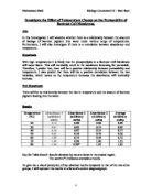
- Table
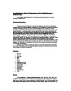
- Graph – attachment at back
- Discussion
-
Analysis of data – looking at my graph I can see as the temperature increases the amount of pigment is released from the cell. But this only begins to happen between 40º and 50º (probably around 45º) before this the raise in temperature seems to have no effect on the membrane. Between 45º and around 60º there is a steady increase in the amount of pigment released. After 60º there is a very significant steep increase in the amount of pigment being released till 70º then there seems to be a decrease in the amount of pigment released.
- Anomalies
Looking at my graph I can see one point that looks like it has to be an anomaly. This would be the last point at 70º. I think it is an anomaly, as it would not make sense for the amount of the pigment in the surrounding cell to decrease. If it had begun to plateau then this could be equilibrium being reached but it actually decreases. Therefore I will disregard this point.
- Conclusion
The data found does support the hypothesis and prediction. As the temperature was increased the amount of pigment released increased therefore showing that some effect has been made on the membrane. My results show the amount of light absorbed from the solution, the higher the number the more absorbed. The more absorbed the darker the colour the pigment is therefore the more released from the cell. For example at 50º the absorption of the colorimeter was just 3.5 but at 60º it was 13 and at 70º it was 164. These results also show that there was a massive increase between 60º and 70º suggesting that something considerable happened between these temperatures. Also the fact that the temperature had no effect on the membrane till approximately 45º is also significant. I have also worked out the correlation coefficient of my graph and found it to be 0.8. This showing at quite strong positive correlation showing that my results follow a general trend.
- Explanation
These results can be explained. I think the reason that there was no change in the membrane till approximately 45º is because at this temperature there is no effect on the components of the cell. Then at 45º we see a slight increase in the amount pigment released and this steadily increases till about 65º. I think the reason for this increase is the beginning of the breakdown of the lipids, creating little holes in the membrane. Then at 65º we see a massive increase in the amount of pigment released. At this point I think it is the protein denaturing causing bigger holes in the membrane allowing the pigment to flow freely through the membrane. I also have to consider that somewhere the fluidity of the membrane would cause holes in the membrane but looking at my graph it is hard to say where this occurs. But looking at my graph it is easy to see that these proteins in the membrane denature at around 65º. There is also the point that the higher the temperature the faster the particles will diffuse anyway. But in this case they diffuse by facilitated diffusion, as they are water-soluble. This means that they require the use of transport proteins and these will become denatured when the temperature is high enough. Looking at my graph I can see no evidence of the graph beginning to plateau so there is no evidence of equilibrium being reached. This may be because my last result is an anomaly.
- Evaluation
Looking at my graph and my correlation coefficient of 0.8 I think it is able to be a general trend therefore proving that my results are good enough to make accurate conclusions. Also the fact that I carried the experiment out of two samples and found an average makes the results more reliable. But I do think that the experiment had many flaws and limitations for the results to be thought of as very reliable. For example the fact that a got an anomalous result which could have told us a lot more about what happened to the cell membrane may have happened because of many errors throughout the experiment.
For example I think one important limitation was the source and condition of the beetroot. It is possible for some of the beetroot to have came from different beetroots each may have been kept in different conditions and may be of different ages all effecting the amount of pigment inside the cells. It should have been made sure that all the beetroot was the same age and had been keep in the same conditions during growth and afterwards. If there were more pigment inside some cells than others then this would have increased the amount diffused.
The size of each slice of beetroot would also be very significant it terms of surface area. We used as tool to get cylinders of beetroot the same diameter but the width was a problem. It is obvious the greater the surface area of the discs the greater chance of diffusion for the pigment. I keep tried to keep the slices at a constant width of 2mm thick but the only equipment available was a scalpel and a ruler so it may not have been very accurate. I better way of cutting them would have been to use a ……….(need to find out) to accurately cut all the beetroot into exact discs of the same width and diameter.
Also the pigment disruption of pigment in the beetroot may be different. The outer surface cells of the beetroot may have more pigment inside them. As the discs of beetroot where from all over the cell it is impossible to say whether they all have the same amount of pigment. To solve this the discs should have be from the same area of the beetroot. If one cell had more pigment in then they will obviously have more pigment to diffuse.
Also the condition the beetroot is kept in during the experiment is important. During the experiment I had my beetroot in an empty beaker. By leaving them in the open air the discs may have become dehydrated meaning when placed into the water the water will first have to enter the cell by osmosis because the water potential inside the cell may be lower than that of the surroundings, this must happen first before the pigments can start to diffuse out, so for some of the time the beetroot is immersed in the water it is re-hydrating. Of course this means the beetroot that was left out the longest will have been the most dehydrated, therefore it would not start to diffuse for a longer time. The modification I would have made then was keeping the beetroot immersed in water during the experiment.
Then there was the limitation of keeping the water bath at a constant temperature with just a Bunsen burner and the use of cold water. I don’t think this would have been a major limitation because as long as the temperature was keep within in a degree or two no major difference would be found, but if it was raised or lowered it could have stopped or started to effect the membrane when not wanted. But a good modification would be the use of an electronic water bath.