Transcription Factors. The promoter is a sequence of DNA near the 5’ end of the coding region where RNA polymerase begins transcription. In prokaryotes there are two essential regions: One, about 40 bp upstream of the initiation point of transcription, is the sequence recognized by RNA polymerase. The second, nearer to the initiation point is rich in AT base pairs (called the TATA box) and is the site where DNA begins to denature so that it’s templates can be exposed. Eukaryotes have a TATA box about 25 bp upstream from the initiation site, and one or two recognition sequences about 50 to 70bp 5’ from the TATA box.
As said before the polymerase cannot simply bind to the DNA promoter and start transcription, it needs some proteins to bind to the DNA before it can bind itself. These proteins are known as Transcription Factors, and with the RNA polymerase make up the Initiation Complex. This binding event between the transcription factors and the DNA presents a new surface for the polymerase to bind to. Regulation can occur here in the way the proteins bind to specific parts, and therefore locations, of DNA. There are currently 4 known methods for proteins to bind to DNA, known as Motifs.
Helix turn helix. This motif occurs in the transcription factors that stimulate specific genes during development and in the proteins that regulate development of the immune and central nervous system.
Zinc finger. Occurs in transcription factors (TF) most notably the steroid hormone receptors.
Leucine zipper. Occurs in many types of DNA binding protein, most notably the inducer AP-1, which binds near promoters of genes involved in mammalian cell growth and division.
Helix loop helix. Occurs in activator proteins that bind to enhancers for the immunoglobulin genes that synthesise antibodies.
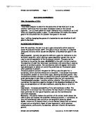
The creation of the initiation complex is shown in the diagram below;
Life, the science of Biology
Firstly transcription factor II D (TFIID) binds to the DNA sequence, using a structure explained shortly, this allows for TFIIA to bind, which prevents any inhibitor proteins binding to the DNA. This is followed by subsequent letters binding to the complex until TFIIH binds. This uses ATP to unwind the DNA helix and allow the polymerase to insert itself in-between. Following that it is postulated that the transcription factor has a protein kinase ability in which it can hydrolyse ATP, transferring the phosphates to multiple serine’s in the c-terminal repeat domain of the largest RNA polymerase subnit. This is thought to release polymerase from the complex and initiate transcription.
However this is only a generalisation of the process as mentioned before the eukaryotes express great diversity in their promoters. The TATA box is common to many promoters but there are some sequences that are specific to only a few genes and are recognised by only a few transcription factors found only in certain tissues. These specific transcription factors play an important role in differentiation. However the TF’s are not degenerate for a specific gene, e.g. TTR (transthyretin) is expressed in liver cells and chloroid plexus but the TF’s used to express it are not all the same.
In addition to transcription factors controlling the regulation of transcription to other regions of DNA bind proteins that activate RNA polymerase. These are the recently discovered regulator regions and enhancer regions. They are situated just upstream and up to 20 kbp away respectively. Regulator proteins bind to the regulator site and their net effect is to bind to the adjacent initiation complex and activate it. Enhancer regions bind activator proteins that strongly stimulate the transcription complex. How they excerpt such an influence from so far away is unclear but in one model the DNA loops around, and the enhancer protein binds back to the transcription complex. However some regulatory proteins can act as repressors by binding to DNA near the promoter site, obscuring it from the transcription factors. Or it can act conversely to activator proteins binding to sites upstream and disrupting the complex.
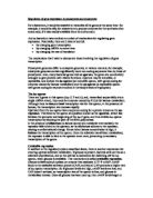
A diagram of the position of the elements in the transcription complex:
Unattributed Internet site.
How do all these different elements actually regulate transcription? The answer seems that all genes in most tissues can transcribe a small amount of RNA. But the right combination of all the above factors determines the maximum rate of transcription. For example in the immature red blood cells of bone marrow, which make a large amount of the protein β-globin, the transcription of globin genes is stimulated by the binding of seven inducers and six activators. By contrast in white blood cells, in the same bone marrow, these regulatory proteins are not made and they do not bind to their sites adjacent to the β-globin genes; consequently these genes are hardly transcribed at all.
The Binding Of Proteins To DNA. The complexity of regulation however starts even before the creation of the initiation complex. The activation of transcription factors is the first step. This can be done in two ways, depending on the type of gene expressed.
Control via lipid soluble hormones. This is gene regulation on an extra cellular level. Lipid soluble hormones, such as steroids are created by other cells and packaged off into the bloodstream. Because they are non-polar molecules they are able to pass through the target cells membrane and into its cytoplasm. Once inside they bind to certain TFs via zinc fingers (explained shortly) to form steroid hormone receptors, which bind to hormone response elements near promoters to activate transcription. For example this occurs in hormone dependent gene activation. The receptor, used to bind the TF’s to the DNA, is in a complex with an inhibitor. Once the hormone enters the cell it out competes the inhibitor for the binding site on the receptor and forms the steroid hormone receptor.
Control via polypeptide hormones. Yet another extra cellular control, but due to the molecule’s polar nature it is unable to pass through the membrane and so binds to a specific receptor on the membrane. Once bound this sets off a cascade reaction in the cell causing the TFs to bind to the DNA. This is referred to as signal transduction, and usually involves phosphorylation.
Once the TFs have been signalled to bind to the DNA the next step is to actually gain access to the gene concerned. Eukaryotic cells have many levels for regulating access.
Genes can be inactivated by chromatin structure. As DNA is packaged up with nuclear proteins it can make genes very inaccessible to the transcriptional apparatus, much like the binding of the repressor to the Lac operon in E-coli. Within a nucleus there are two types of chromatin, the DNA protein complex, Euchromatin and Heterochromatin. Euchromatin is diffuse and is transcribed into mRNA. Heterochromatin is denser and is generally not transcribed. This is best described with the example of the X chromosome in mammals. The female of the species has the potential to produce twice as much gene products as the male (The Y chromosome in most animals is largely transcriptionally inactive). If this mutation of double expression were on an autosome it would usually be lethal and the embryo would fail to develop. So how do both sexes develop when one of them obviously has an extra, or one less, chromosome.
The answer is that by random choice in the female every cell has an inactive condensed X chromosome, comprising of heterochromatin, known as the Barr Body. This inactivation of the chromosome appears to be brought about by Cytosine Methylation. This methylation prevents TFs binding due to certain chromosomal proteins binding in place.
Even when the correct part of DNA is accessible the transcription factors have to cope with the histone proteins present in the nucelosomes, which could block TFs from binding. Several mechanisms are employed by the eukaryotic cell to help prevent this. For example the TFs bind just after DNA replication before the histones have a chance to bind or molecules known as nucleosome disruptors open up the nucleosome complex and allow the TFs to bind.
In some cases gene expression is regulated by movement of a gene to a new location on the chromosome. This occurs in the yeast Sacchromyces cerevisiae with respect to it haploid mating type. The haploid cell can be either a or α depending on the translocation of the genes to a third site known as the MAT locus. Whenever the gene gets moved to this region it is expressed, whilst the other is not.
Regulation can also be controlled regarding the number of genes coding for a particular protein that are present. The process of creating more genes to increase transcription is known as Gene Amplification. We see this process occurring in synthesis of rRNA of higher eukaryotes. The mechanism for selective gene overreplication is not fully understood.
Posttranscriptional Control
The number of methods for regulating gene expression are reduced here in comparison although they allow for very interesting manipulation of the final product, rather than controlling the rate of its production.
RNA Processing. The primary transcript of the RNA obtained from the DNA contains many introns (non coding sections of DNA) unlike that of the prokaryotes, which is suitable for translation as soon as it leaves the polymerase. These introns are usually removed in the conversion of the pre-mRNA to the mature mRNA which goes to the ribosomes. The action of cutting out the introns leads to a very interesting form of regulation. Suppose a primary transcript had two introns and during splicing something went wrong and the start of the first intron was spliced to the end of the second. No only would you remove the two introns but also the middle exon. This would lead to a radically different protein being produced. Although alternative splicing is uncommon in normal RNA processing it can be used to create a whole family of proteins from a single gene. For example the primary transcript for tropomyosin, with it’s 11 introns can be alternately spliced to give 5 different proteins.
Some viral proteins can also regulate the process of moving the mRNA out of the nucleus to the cytoplasm, although examples of this are rare. The Adenovirus mRNAs have priority over all cellular mRNA when moving into or out of the nucleus. This suggests that it is not specific to a certain type of gene although the reasons as to why this is are uncertain.
Also the stability of the mRNA can be regulated. DNA has to be stable and has many complex repair mechanisms to keep it that way. However RNA doesn’t have this luxury, when it gets out into the cytoplasm is subject to attack by ribonucleases and lysosomes. Eventually all eukaryotic mRNA degrades but the time it take can affect the amount of protein it is able to express. It has been found that the half-life of a strand of mRNA is directly proportional to the length of a repeating AUUUA sequence on the 3’ untranslated part of the mRNA. The mechanism by which these sequences stabilise the strand is not yet known.
Translational and Posttranslational Control
Once the mRNA has been cut down to the appropriate size it is ready to code for a protein by attaching itself to a ribosome. However the amount of free mRNA in the cell can be regulated by binding it to a protein in a negative feedback fashion in some cases to stop a cell overproducing a certain protein. For example the translational repressor protein that binds to ferritin mRNA when iron levels are too high in the blood.
The process of translation can be controlled. One method of control is the use of capping at the 5’ end of the mRNA, via a modified guanosine residue. mRNA’s that do not have a modified cap are not translated. This modification of the normal guanosine cap can occur at any time during development making the mRNA active.
The final method for control in gene expression regards the modification of proteins after synthesis, such as glycosylation or phosphorylation. Also their lifespan in the cell is important. For example transcriptional inducers must only be present when needed otherwise the gene would always be on. A small protein called ubiquitin is used in the breakdown of proteins. It collects around the surface of the protein and attracts a large complex of proteases which catalyse its breakdown.
Even single celled organisms such as yeasts have many of these complex regulation mechanisms. In multicellular organisms these mechanisms must also co-ordinate the activities of different cells and tissues. Gene regulation is all about the expression of genotype into phenotype. This is most impressive when looking at the development of a single cell into a complex organism.
Bibliography
Alberts (1994) Molecular Biology of the Cell 3rd Edition
Lodish (1995) Molecular Cell Biology 3rd Edition
Purves (1998) Life The Science of Biology 5th Edition
Various Internet sites found from a google search on “mechanisms controlling eukaryotic gene expression”