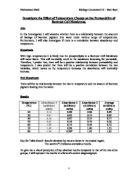
RESULTS
Table of results:
Results in bold: anomalous results identified.
TRENDS AND PATTERNS
Looking at the graph, it can be seen that as the temperature increased, the absorbance of the solution around beetroot also increased.
Between 30-40°c, there is a significantly small increase in the absorbance of my results but there is a slight decrease in the mean results.
Between 40-50°c, the opposite happens here, my results show that the absorbance decreases but the mean results show that the absorbance increases
Between 50-60°c, there was an increase in the absorbance
Between 60-70°c, there was a very steep increase in the absorbance
Between 70-80°c, there was an increase in both results
The difference in the results at the beginning of the graph was probably due to anomalous results.
CONCLUSION
The graph clearly shows that as the temperature increases, the absorbency also increases.
The beetroot discs were washed in the experiment to remove any anthocyonin from the surface. The colour of the liquid in the test tubes changed due to diffusion taking place. Anthocyonin diffused out from the cell into the liquid. This happened because of the big concentration gradient. The results showed that as the temperature was increased the membrane became more permeable and allowed more pigment to enter the liquid from the beetroot.
DISCUSSION
The cell membrane contains intrinsic proteins that control the membrane’s selective permeability. There are not any channels for anthocyonin to come through, as it is not supposed to come out of the cell. It only diffuses out as the proteins in the membrane become denatured. The structure is destroyed as the heat provides them with kinetic energy and they make space between them, this causes the membrane to become selectively permeable.
Protein structure has a hierarchical classification. The first order of structure, the primary (1°) structure, is the type and sequence of the (covalently bonded) amino acids that constitute the polypeptide chain from which the mature protein is derived. Secondary (2°) structure describes the localised folding patterns adopted by discrete regions or domains within the polypeptide chain e.g. the a-helix or ß-sheet. Tertiary (3°) structure is the overall shape of a globular protein and is determined by the way in which the whole polypeptide chain folds. Tertiary structure is maintained by inter- amino acid interaction involving e.g. hydrophobic interactions and disulphide bonds; such interactions may involve amino acid residues that are linearly distant yet spatially proximal. Some polypeptides having folded to their correct tertiary structure may bond with other sub-unit polypeptides during assembly of a protein, so producing a quaternary (4°) structure. This structure accounts for the cell letting the anthocyonin out.
EVALUATION
Errors:
- Some beetroot may have more anthocyonin than others. This is trivial though; it would not make much difference.
- The beetroot discs were not measured as accurately as they seemed. They could be significantly too large or small.
- The equipment used was not accurate enough. Using a pipette is not necessarily accurate; it could have produced errors in measurement.
Limitations:
- The whole experiment should be repeated more times to make the results more reliable
- Only 6 temperatures were looked at, we could have done more
- When the beetroot was stabbed to put on the needle, it could have allowed anthocyonin to come out. But this was done so that the beetroot discs don’t touch one another and maximum surface area is created.
- Errors could have been made when using the colorimeter, it as a machine and they can make accuracy errors.
- Cuvettes had to be handled very carefully, but scratches may have already been on then and fingerprints also affect the absorbency. So in effect, these could have taken place.