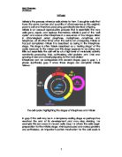
Interphase can be categorized into several stages, gap 0, gap 1, s phase (synthesis), gap 2 once these stages are complete mitosis follows.
The cell cycle: highlighting the stages of interphase and mitosis
In gap 0 the cell may be in a temporary resting stage or perhaps has reached the end of its development and may stop dividing. For example this can occur in neuron cells. Gap 1 is where the cells begin preparation for the mitotic stage. RNA is produced and various proteins are synthesized. An important control mechanism for the cell cycle is activated in this stage, ensuring everything is ready for DNA synthesis. DNA to be replicated is synthesised during this stage of Interphase. Before entering mitosis, the cell endures one final stage of Interphase before continuing, in gap 2 the cell continues to grow and perform its synthesis of proteins. At the end of this occurs another control checkpoint to determine whether or not the cell can enter mitosis and divide to produce to daughter cells.
Following Interphase the cell enters the first stage of the mitotic process, known as prophase. The Process as whole is much shorter in comparison to Interphase and by this stage much of the cells common functions have stopped and attention within the cell is centred on the division. In prophase chromosomes become visible; the nucleolus fades and chromatin (replicated DNA and associated proteins) condense to form chromosomes. Chromosomes consist of two chromatids comprising of the same genetic information repeated twice in coils of DNA and centromeres -the central point on the chromosomes that connects each chromatid together to form a chromosome.
Centrioles that divided during Interphase form spindle fibres long chains of microtubules that ‘latch’ onto each centromere. There is a period sometimes called prometaphase, which is when the nucleus ceases to exist and the nuclear envelope breaks down. Some mitotic spindle fibers elongate from the centrioles and attach to kinetichores, protein bundles located on the chromosomes. Other spindle fibers elongate but instead of attaching to chromosomes, overlap each other at the cell centre or equator.
Metaphase follows with the spindle fibres attaching to the chromosomes and their tension through attachment causes the chromosomes to forcefully align on the equator of the cell.
The centromeres split under the tension and spindle fibres shorten causing the chromatids of each chromosome to separate and migrate to opposite poles making daughter chromosomes. This is Anaphase. The shortening of the spindle fibres is caused the removal of tubulin the very molecules the fibres are comprised of.
The final stage of mitosis is telophase where the daughter chromosomes reach each pole of the cell and uncoil with the reformation of a nuclear envelope forming around each bundle. They now become ‘invisible’ again and the spindle fibres disintegrate, and a nucleolus reforms in each new nucleus. Cytokinesis, the actual division of the cell can be preformed by most animal cells can perform mitosis however only a specialised group of cells can do so in plants these are known as meristematic cells
In animal cells cytokinesis occurs by the constriction of the centre of the parent cell from outside inwards. In plant cells this differs by the growth of a cell plate that forms across the equator of the cell. It is formed from the vesicles produced by the dictyosome or golgi apparatus. Cellulose then form over this plate to create the beginnings of a cell wall a characteristic that is quite obviously absent in animal cells.