Sperm Production
Each testis is a mass of more than 800 tightly looped and folded vessels known as Seminifeous tubules. Inside each tubule, sperm begin as blob-like cells called Spermatogona lining the inner wall. These pass through larger stage, as Primary Spermatocytes, and begin to develop tails as Spermatids. As all of this happens, they move steadily towards the middle of the tubule. The spermatids finally develop into ripe sperm with long tails. Thousands of sperm are produced every second, each taking about two months to mature.
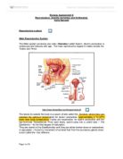
Female Reproductive System
Unlike the male, the female reproductive organs are sited entirely inside the body.
Reproductive tract
The female reproductive glands Ovaries are located within the abdomen. From puberty, they mature and release the female sex cells (gametes), known as egg or ova. This release occurs in average once a month as part of the menstrual cycle. The ripe egg travels along the fallopian tube to the uterus, the muscular sac in which it develops into an Embryo and then a Foetus. Unfertilised eggs and the uterus lining leave via the vagina. The ovaries also make the female sex hormone Oestrogen.
Ovulation
An ovary contains thousands of immature egg cells. During each menstrual cycle, follicle-stimulating hormone (FSH) causes one egg to begin development: this takes place inside a Primary Follicle. The follicle enlarges as its cell proliferates, and begins to fill with fluid, becoming a Secondary Follicle that moves to the ovary’s surface. It also increases its production of the hormone oestrogen. A surge of Luteinizing Hormone (LH) causes the follicle to rupture and release the ripe egg – this is Ovulation. The lining of the empty follicle thickens into a Corpus- Luteum.
http://cache-media.britannica.com.cdnetworks.net//eb-media/36/99236-004-770A8041.jpg
Fertilisation
Fertilisation is the term used to describe the exact moment when the Nuclei of the male and female gametes fuse. Each gamete contains half the full number of chromosomes, fertilisation creates a new cell with the full number of chromosomes this cell is called a Zygote. The Zygote contains two sets of chromosomes – one set from the male parent and the other from the female parent.
Combining the genetic materiel from the two individuals makes offspring that are genetically Unique.
http://members.optushome.com.au/karens/fertilisation.gif
In humans, sperm are deposited high up in the female vagina close to the entrance of the cervix. Once there, they have to make their way up through the cervix and uterus, and into one of the oviducts. Once the sperm are in the oviduct fertilisation may occur.
- The sperm swim towards the egg cell in the oviduct.
- Once one sperm makes contact with the zona pellucida of the egg cell the acrosome reaction occurs- this is where digestive enzymes released from the acrosome of the sperm.
- These enzymes digest the zona pellucida, so that the sperm can move through it to the plasma membrane of the egg cell.
- The sperm head fuses with the cell membrane of the egg cell. This triggers the cortical reaction-the egg cell releases the contents of vesicles called cortical granules into the space between the cell membrane and the zona pellucida.
-
The chemicals from the cortical granules make the zona pellucida thicken, which makes it impenetrable to the other sperm. This makes sure that only one sperm fertilises the egg cell.
- Only the sperm nucleus enters the egg cell- its tail is discarded.
- The nucleus of the sperm fuses with the nucleus of the egg cell- this is fertilisation.
A Zygote is now formed, which has the full number of chromosomes. It immediately begins to divide by Mitosis to develop into a fully formed offspring.
Stages of Implantation
Fertilisation
Fertilisation takes place in the fallopian tube when the head of a sperm cell, or spermatozoon, penetrates the egg. This forms a single cell (Zygote).
http://www.bio.davidson.edu/Courses/Molbio/MolStudents/spring2005/Champaloux/fertilization.jpg
Zygote
The fertilised egg passes along the fallopian tube. Within 24-36 hours it has divided into two cells, then 12 hours later into four cells, and so on. This process is known as cleavage.
http://sprott.physics.wisc.edu/pickover/zygote.jpg
Morula
The zygote divides several times to form a solid blackberry- like cluster of 16-32 cells, the morula (derived from the Latin for “Mulberry”). At 3-4 days after fertilisation the morula leaves the fallopian tube and enters the uterine cavity.
http://blogs.discovermagazine.com/loom/files/prevsite/morula.gif
Blastocyst
About six days after fertilisation, the cell cluster forms a hollow cavity and is known as a Blastocyst. It floats within the uterus for around 48 hours before landing on the thick uterus lining (Endometrium), which softens to aid implantation (Burrowing of the Blastocyst into the Endometrium). The inner group of cells become the embryo itself.
http://www.molecularstation.com/molecular-biology-images/data//504/thumbs/Blastocyst.jpg
Embryonic disc
Within the inner cell mass, an Embryonic disc forms. This separates the cell cluster into the amniotic cavity, which develops a sac that fills with fluid and fold around to cover the embryo, and the yolk sac, which helps to transport nutrients to the embryo during the second and third weeks. The disc develops three circular sheets called the primary germ layers-ectoderm, mesoderm and endoderm – from which all body structures will derive.
http://cache-media.britannica.com.cdnetworks.net//eb-media/79/1079-004-38FAAFCC.gif