-
Bladder: Two ureters track down the back of the abdomen and feed into the bladder. An average bladder can normally hold about half a pint of urine before the brain tells the person they need to excrete it. If ignored then the bladder can expand to hold around a pint of urine. Yet the bladder can still stretch if necessary, as happens in disorders involving retention of urine.The bladder’s inner lining is formed from a folded, wrinkled membrane. As the bladder expands this membrane flattens out to maintain its barrier.
-
Ureters: These are two long tubes that collect the urine from the pelvis and run it down the back of the abdomen and feed into the bladder via two small flap like openings
-
Urethra: This is one short tube which extends from the bottom of your bladder and expels your urine from the body.
Kidney defects or diseases
The kidneys are organs which can be easily damaged. They are only protected by a small amount of fatty tissue around them which can make them an easy target area. Also they are susceptible to diseases which can make them need help to work or completely fail. But these diseases can be controlled or overcome with different methods and different medicines. Below are some of the diseases which can affect the kidneys.
- Nephritis
The worst form of nephritis is glomerulonephritis. This condition affects children and teenagers far more often than it affects adults. It is inflammation of the glomeruli.
Acute glomerulonephritis usually develops a few weeks after an infection of the throat or skin. Symptoms of glomerulonephritis include fatigue, high blood pressure, and swelling. Swelling is most notable in the hands, feet, ankles and face.
Pyelonephritis (an adults disease) usually occurs suddenly, and the acute form of this disease is more common in adult women. The most common cause of this form of bacterial nephritis is the backward flow of infected urine from the bladder into the upper urinary tract. Its symptoms include fever and chills, fatigue, burning or frequent urination, cloudy or bloody urine, and aching pain on one of both sides of the lower back or abdomen.
Diagnosis of nephritis is based on:
- The patient's symptoms and medical history
- Physical examination
- Laboratory tests
- Kidney function tests
- Imaging studies such as ultrasound or x rays to determine blockage and inflammation..
- Diabetes.
Diabetes means that your blood glucose (often called blood sugar) is too high. Your blood always has some glucose in it because your body needs glucose for energy to keep you going. But too much glucose in the blood isn't good for your health.
Glucose comes from the food you eat and is also made in your liver and muscles. Your blood carries the glucose to all the cells in your body. Insulin is a chemical (a hormone) made in a part of the body called the pancreas. The pancreas releases insulin into the blood. Insulin helps the glucose from food get into your cells. If your body doesn't make enough insulin or if the insulin doesn't work the way it should, glucose can't get into your cells. It stays in your blood instead. Your blood glucose level then gets too high, causing you to have diabetes.
The signs of diabetes are:
- being very thirsty
- urinating often
- feeling very hungry or tired
- losing weight without trying
- having sores that heal slowly
- having dry, itchy skin
- losing the feeling in your feet or having tingling in your feet
- having blurry eyesight
You may have had one or more of these signs before you found out you had diabetes. Or you may have had no signs at all.
People can get diabetes at any age. There are three main kinds.
Type 1 diabetes, formerly called juvenile diabetes or insulin-dependent diabetes, is usually first diagnosed in children, teenagers, or young adults. In this form of diabetes, the beta cells of the pancreas no longer make insulin because the body's immune system has attacked and destroyed them. Treatment for type 1 diabetes includes taking insulin shots or using an insulin pump, making wise food choices, exercising regularly, taking aspirin daily, and controlling blood pressure and cholesterol.
Type 2 diabetes, formerly called adult-onset diabetes or non-insulin-dependent diabetes, is the most common form of diabetes. People can develop type 2 diabetes at any age -- even during childhood. In type 2 diabetes, the pancreas does not make enough insulin, and the fat, muscle, or liver cells do not use it properly. Being overweight can increase the chances of developing type 2 diabetes. Treatment includes using diabetes medicines, making wise food choices, exercising regularly, taking aspirin daily, and controlling blood pressure and cholesterol.
Gestational Diabetes
some women develop gestational diabetes during the late stages of pregnancy. Although this form of diabetes usually goes away after the baby is born, a woman who has had it is more likely to develop type 2 diabetes later in life. Gestational diabetes is caused by the hormones of pregnancy or a shortage of insulin.
- Hypertension
Hypertension is high blood pressure. Blood pressure is the force of blood pushing against the walls of arteries as it flows through them. Arteries are the blood vessels that carry oxygenated blood from the heart to the body's tissues.
As blood flows through arteries it pushes against the inside of the artery walls. The more pressure the blood exerts on the artery walls, the higher the blood pressure will be. The size of small arteries also affects the blood pressure. When the muscular walls of arteries are relaxed, or dilated, the pressure of the blood flowing through them is lower than when the artery walls narrow, or constrict.
Hypertension is a major health problem, especially because it has no symptoms. Many people have hypertension without knowing it. Hypertension is more common in men than women and in people over the age of 65 than in younger persons. Hypertension is serious because people with the condition have a higher risk for heart disease and other medical problems (such as kidney damage) than people with normal blood pressure. Serious complications can be avoided by getting regular blood pressure checks and treating hypertension as soon as it is diagnosed.
If left untreated, hypertension can lead to the following medical conditions:
- Arteriosclerosis, also called atherosclerosis
- Heart attack
- Stroke
- Enlarged heart
- Kidney damage.
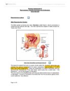
4. Overproductive/underproductive ADH
Description
Antidiuretic hormone (ADH) is an important hormone in maintaining normal water balance. Too little ADH results in diabetes insipidus, which is manifested by large volumes of water in the urine. Too much ADH results in the syndrome of inappropriate antidiuretic hormone secretion (SIADH), with water retention and decreased blood sodium levels.
The diagnosis is established through a combination of blood and urine tests performed under certain specified conditions. The patient must not be dehydrated or volume overloaded. Water restriction is the cornerstone of treatment. Decreased water intake allows the serum sodium level to rise normally. The maximum amount of water that patients are allowed to drink is just slightly more than the amount of urine they produce. Patients must have regular serum sodium measurements to ensure that the water restriction has been effective. Some patients may require a diuretic such as furosemide if further treatment is needed.
The most concerning potential side effect from treatment is dehydration. This occurs when water restriction is maintained in a patient with increased fluid requirements because of fever, exercise, or other reasons.
There is one way in which most kidney diseases or problems can be dealt with if needed. This is kidney transplants, these operations are regularly carried out in the u k and they involve one patient (alive or dead) donating an organ to another patient (recipient). In a living donor transplant, a kidney from a donor, usually a blood relative is transplanted into your body. The most suitable donors are usually members of your immediate family. Sometimes a spouse, distant relative, or close friend can also be a suitable donor.
The donor's blood group and tissue type must be compatible with yours, and extensive medical tests will be done to determine the health of the donor. People who donate a kidney can live a normal life with one kidney and there are few risks to healthy donors. For this type of transplant, there is a shorter waiting period and the transplant operation is planned at a time convenient for you and your donor.
Living donor transplants have a 90 to 95% success rate. That means that after one year, 90 to 95 of every 100 transplanted kidneys are still working. A transplant from a non-living donor is called a cadaveric transplant. In this type of transplant, a healthy kidney from someone who has died suddenly is transplanted into your body. Before a cadaveric donor's organs can be transplanted, a series of medical tests is done to determine if they are healthy. In addition, the family of the donor must consent to organ donation. You will not know the identity of your donor.
After you have a series of tests, you will be put on a transplant waiting list until a kidney is found that is compatible with your body. The length of time you will have to wait is hard to predict because it depends on how hard you are to match and how many kidneys become available. Unfortunately, the waiting time for a cadaveric organ transplant is getting longer. Cadaveric transplants have an 80 to 85% success rate.
The transplant operation usually takes two to four hours. The new kidney and ureter (the tube through which the urine flows into the bladder) are placed in your lower abdomen near the groin. They are surgically attached to your blood vessels and bladder. Your old kidneys are not removed unless they are so large there is no room for the new kidney, or they are chronically infected.
LOCATION OF TRANSPLANTED KIDNEY
Dialysis is a treatment for people in the later stage of chronic renal insufficiency (kidney failure). This treatment cleans the blood and removes wastes and excess water from the body. Normally, this work is done by healthy kidneys.
Sometimes dialysis is a temporary treatment. However, when the loss of kidney function is permanent (as in end-stage kidney failure), you must continue to have dialysis on a regular basis. The only other treatment for kidney failure is a kidney transplant.
There are two types of dialysis: hemodialysis and peritoneal dialysis. In hemodialysis, your blood is passed through an artificial kidney machine to clean it. Peritoneal dialysis uses a filtration process similar to hemodialysis, but the blood is cleaned inside your body rather than in a machine. Hemodialysis means "cleaning the blood"-and that is exactly what this treatment does. Blood is circulated through a machine which contains a dialyzer (also called an artificial kidney). The dialyzer has two spaces separated by a thin membrane. Blood passes on one side of the membrane and dialysis fluid passes on the other. The wastes and excess water pass from the blood through the membrane into the dialysis fluid which is then discarded. The cleaned blood is returned to your bloodstream. You can be attached to the dialysis machine in different ways. The most common method of providing permanent access to the bloodstream for hemodialysis is an internal fistula in your arm. This involves having an artery and a vein connected surgically. When they are joined, the stronger blood flow from the artery causes the vein to become larger. Needles can be inserted in the enlarged vein to connect you to the dialysis machine.Another way to provide access to the bloodstream is to insert an internal graft. In this procedure an artery is surgically connected to a vein with a short piece of special tubing placed under the skin. Needles can be inserted in this graft.
There is another form of dialysis called peritoneal dialysis. Peritoneal dialysis works on the same principle as hemodialysis, but the blood is cleaned inside the body rather than through a machine. Your abdomen has a peritoneal cavity, lined by a thin membrane, called the peritoneum, which surrounds the intestines and other internal organs. The peritoneal cavity in the abdomen is filled with dialysis fluid that enters the body through a permanently implanted catheter. Excess water and wastes pass though the peritoneum into the dialysis fluid. Then, this fluid is drained from the body and discarded. The process is repeated between three and five times a day. In most cases, this treatment can be performed without assistance, at home or at work. A tube called a catheter, made of soft, non-irritating plastic, is inserted in your abdomen below and to one side of your navel, and stays there as long as you are using this type of dialysis. The catheter may be inserted at the bedside using local anesthetic, or in the operating room, depending on what is best for you. The dialysis fluid flows into, and is drained out of, the peritoneal cavity through this special tube. The insertion of the catheter may cause discomfort for a brief period, but peritoneal dialysis is not painful. However, care must be taken to avoid infection.
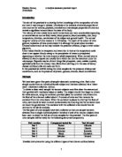
Kidney diseases and defects all have to be diagnosed so that docters and nurses can give them the appropriate treatment for the problem. These can be tested for in a variety of diffent ways. There are different tests for different diseases below is a write up of a tests on different samples of urine for different diseases.
Bibliography.
- A-level biology.
- Advanced biology.
- Diabetes book. (diabetes for dummies)
- as level biology book.