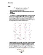
If you would wish to reverse this reaction you would use a hydrolysis reaction. To do this you would add water.
This is an example of a condensation reaction which results in the formation of peptide bonds.
When two amino acids are combined this forms a dipeptide.
When further amino acids are combined this results in the formation of a polypeptide.
The Structure of proteins
The function of a protein is determined by its shape. The shape of a protein is determined by its (sequence of amino acids). The sequence of amino acids in a protein is determined by the sequence of nucleotides in the gene (DNA) encoding it. The function of a protein is absolutely dependent on its three-dimensional structure. changes in (alters between charged amino acids) changes in salt concentration (does the same) changes in temperature (higher temperatures reduce the strength of ) presence of reducing agents (break S-S bonds between )
None of these agents breaks , so the primary structure of a protein remains intact when it is denatured.
When a protein is denatured, it loses its function. Often when a protein has been gently denatured and then is returned to normal physiological conditions of temperature, pH, salt concentration, etc., it spontaneously regains its function (e.g. enzymatic activity or ability to bind its antigen).
Primary Structure
The primary structure of a protein is its linear sequence of amino acids and the location of any disulfide (-S-S-) bridges.
Note the amino terminal or "N-terminal" (NH3+) at one end; carboxyl terminal ("C-terminal") (COO-) at the other.
Secondary Structure
Most proteins contain one or more stretches of amino acids that take on a characteristic structure in 3-D space. The most common of these are the alpha helix and the beta conformation.
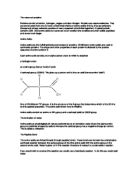
Alpha Helix
-
The all extend to the outside.
- The helix makes a complete turn every 3.6 amino acids.
- The helix is right-handed; it twists in a clockwise direction.
-
The carbonyl group (-C=O) of each extends parallel to the axis of the helix and points directly at the -N-H group of the peptide bond 4 amino acids below it in the helix. A forms between them
[-N-H·····O=C-] .
Beta Conformation
- consists of pairs of chains lying side-by-side and
- stabilized by hydrogen bonds between the carbonyl oxygen atom on one chain and the -NH group on the adjacent chain.
-
The chains are often "anti-parallel"; the direction of one being the reverse of the other.
Tertiary Structure
Tertiary structure refers to the three-dimensional structure of the entire .
Where the entire protein or parts of a protein are exposed to water (e.g., in blood or the cytosol), hydrophilic — including R groups with sugars attached — are found at the surface; hydrophobic R groups are buried in the interior.
The function of a protein depends on its tertiary structure. If this is disrupted, the protein is said to be denatured
A in the gene encoding a protein is a frequent cause of altered tertiary structure.
-
The mutant versions of proteins may fail to reach their in the cell and/or be degraded.
Examples:
- Most cases of cystic fibrosis
-
is caused by improper folding of mutant versions of
-
V2 - the (ADH) receptor or
Mutant proteins may aggregate forming insoluble, nonfunctional deposits. This is particularly likely if the mutation causes to be displayed at the surface of the molecule rather than in its interior.
The tertiary structure of many proteins is built from several domains.
Often each domain has a separate function to perform for the protein, such as:
- binding a small ligand (e.g., a peptide in the molecule shown here)
-
spanning the plasma membrane ()
-
containing the catalytic site ()
-
DNA-binding (in )
-
providing a surface to bind specifically to another protein []
Quaternary Structure
Complexes of 2 or more held together by but in precise ratios and with a precise 3-D configuration.
The noncovalent association of a molecule of beta-2 microglobulin with the heavy chain of each is an example.
Collagens
Collagens are
-
insoluble, extracellular
- found in all animals
- the most abundant proteins in the human body
They are essential structural components of all connective tissues, such as
- cartilage
- bone
- tendons
- ligaments
- fascia
- skin
19 types of collagens have been found (so far) in humans. The major ones are:
- Type I. The chief component of tendons, ligaments, and bones.
-
Type II. Represents more than 50% of the protein in cartilage. It is also used to build the of vertebrate embryos.
-
Type III. Strengthens the walls of hollow structures like arteries, the intestine, and the uterus.
-
Type IV. Forms the basal lamina of epithelia. (The basal lamina is often called the basement membrane, but is not related to lipid bilayer membranes.) A meshwork of Type IV collagens provides the filter for the blood capillaries and the of the kidneys.
The other 15 types are probably equally important but they are much less abundant.
Primary Structure of Collagens
The basic unit of collagens is a polypeptide consisting of the repeating sequence
( (Gly) - X - Y)n
where X is often (Pro) and Y is often hydroxyproline (proline to which an -OH group is added after synthesis of the polypeptide).
Secondary and Tertiary Structure
The resulting molecule twists into an elongated, left-handed helix (NOT an ). When synthesized, the and C- terminal of the polypeptide have globular domains, which keep the molecule soluble.
As they pass through the endoplasmic reticulum (ER) and ,
- The molecules are glycosylated.
- Hydroxyl (-OH) groups are added to the "Y" amino acid.
- S-S bonds link three chains covalently.
- The three molecules twist together to form a triple helix.
In some collagens (e.g., Type II), the three molecules are identical (the product of a single gene). In other collagens (e.g., Type I), two polypeptides of one kind (gene product) assemble with a second, quite similar, polypeptide, that is the product of a second gene.
When the triple helix is secreted from the cell (usually by a fibroblast), the globular ends are cleaved off. The resulting linear, insoluble molecules assemble into collagen fibers. They assemble in a staggered pattern that gives rise to the striations seen in this electron micrograph (courtesy of Dr. Jerome Gross). (Type IV collagens are an exception; they form a meshwork rather than striated fibers.)