Reading of results
The results of Microbiological examinations usually become available in stages on successive days. Microscopial observations on stained films may be obtained on the day of receipt of the specimen and if significant, be given in a preliminary report to the clinician.
The results of these later tests are generally available on the third day, when the content of the final report can be decided. For some types of examination, for example that of the urine for significant bacteriuria, diarrhoeal faeces for enteropathogens or sera for specific antibodies, the sequence of test procedures, the criteria for reading the results and the phrasing to be used in reports can be clearly defined in a manual of instructions.
When many examinations have to be made, even skilled and conscientious workers may from time to time make mistakes. Staff should be encouraged to recognise and report any likelihood of their having made a mistake, and should not be made afraid to confess the possibility.
Wording of reports
The aim of the clinical microbiologist is to provide clinicians and health officers with reports that are understandable, instructive and relevant as well as reliable. The laboratory should therefore have a carefully considered policy for the wording of reports and all staff should adhere to that policy.
The laboratory‘s for reports should specify not only the wording of interpretative comments, but also circumstances in which the different comments are to be made. A policy is also required for reporting the finding of acid-fast bacilli in different specimens. Thus, their finding in sputum might be reported, ‘Acid fast bacilli resembling tubercle bacilli seen in film.
Particular care must be given to the policy for the wording of negative reports. These should be phrased in such a way as to indicate which pathogens were sought and not found. They should not suggest that tests had been made for a wide range of pathogens when indeed methods for detecting only a few types of pathogenic bacteria had been used.
Laboratory Manual
A prime responsibility of the director of a laboratory is to compile or supervise the compilation of a laboratory manual of procedures, comprising a collection of instruction sheets for the different sections of the laboratory. The manual should lay down the policy of the laboratory for the kinds and sequence of examinations to be made on each of the different kinds of specimen, the criteria for determining the content of reports, and the standardised wording of reports.
Cholera Disease
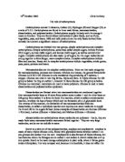
The Features of Cholera
As the disease is water-borne, it occurs where people do not have access to proper sanitation, a clean water supply or uncontaminated food. Infected people, three-quarters of whom may be symptomless carriers, pass out large numbers of bacteria in their faeces. If these contaminate the water supply, or infected people handle food or cooking utensils without washing their hands, then bacteria are transmitted to uninfected people.
Almost all people with cholera who are treated make a quick recovery. A death from cholera is an avoidable death.
The figure below shows an electron micrograph of Vibrio cholerae. The faeces of a cholera victim are full of these bacteria with their distinctive flagella.
Malaria Disease
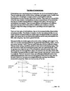
The Features of Malaria
Malaria is still one of the world’s biggest threats to health. 40% of the world’s population live in areas where there is a risk of malaria.
The figure below shows the life cycle of Plasmodium. The parasite has two hosts: the sexual stage occurs in mosquitoes, the asexual stage in humans. The time between infection and appearance of parasites inside red blood cells is 7-30 days in P.falciparum; longer in other species.
HIV (Human Immunodeficiency Virus) Disease
The features of HIV
Infection with HIV occurs by the transfer of , , , , or . Within these bodily fluids, HIV is present as both free virus particles and virus within infected immune cells. The four major routes of transmission are , contaminated needles, breast milk, and transmission from an infected mother to her baby at . Screening of blood products for HIV has largely eliminated transmission through blood transfusions or infected blood products in the .
Tuberculosis (TB) Disease
The features of HIV
Tb is caused by two bacteria, Mycobacterium tuberculosis and M. bovis. These are pathogens that live inside the human cells, particularly in the lungs. This is the first site of the infection, but the bacteria can spread throughout the whole body and even infect the bone tissue. Some people become infected and develop TB quite quickly, whilst in others the bacteria remain inactive for many years.
It is estimated that 30% of the world’s population is infected with TB without showing any symptoms of infection; people with this inactive infection do not spread the disease to others.
Equipment used in the Microbiology Department
it is convenient and time-saving to have most of the commonly used equipment such as incubators, refrigerators, microscopes and water baths located in the laboratories where the main work is done rather than in separate instrument rooms. Exceptions are noisy machines such as centrifuges, rotary incubators and shakers, which are best housed in a separate room.
Of all the equipment used in clinical microbiology, most careful consideration is required in the choice of autoclaves. Different conditions of autoclaving are required for different kinds of materials, e.g. discarded cultures, clean glassware and large volumes of culture media, and it is necessary to have a modern multi-purpose machine that can readily and reliably be switched to different cycles of operation or to have different autoclaves for each different function.
Expensive, delicate and elaborate equipment is now becoming available to perform rapidly and automatically tests that are slow and sometime inexact by conventional methods. Any decision to install such equipment should be influenced by considerations of cost effectiveness and clinical value and must take account of the will and ability of staff to maintain the equipment in good working order.
Safety Precautions
Every clinical microbiology laboratory must have a defined system for instructing and supervising staff in safety precautions. Whilst the head of the laboratory bears the ultimate responsibility for the safety arrangements, another senior member of staff may be appointed to act as safety officer to coordinate the arrangements and supervise their implementation. All staff should be offered appropriate immunizations, e.g. that against tuberculosis, and some staff should be trained in first aid and have ready access to basic first aid equipment.
Inoculation loop
An inoculation loop (sometimes called a smear loop, inoculation wand or microstreaker) is a simple tool used mainly by to retrieve an from a of microorganisms. Its tip is a wire made of or , the latter being inferior but less expensive. The wire forms a small loop with a diameter of about 5 mm. This loop is handy for taking an inoculum from a liquid by using the phenomenon of .
The inoculation loop is always in a flame until it becomes red hot before and after each use. By doing this, the same tool can be reused in different experiments without fear of cross-contamination.The loop is used to cultivate microbes in Agar jelly and to use the streak manouvre to transfer microbes.
Incubator (microbiology)
In microbiology, an incubator is a device for controlling the temperature, humidity, and other conditions in which a is being grown. The simplest incubators are insulated boxes with an adjustable heater, typically going up to 60 to 65 °C (140 to 150 °F), though some can go slightly higher (generally to no more than 100 °C). More elaborate incubators can also include the ability to lower the temperature (via refrigeration), or the ability to control humidity or levels.
Most incubators include a timer; some can also be programmed to cycle through different temperatures, humidity levels, etc. Incubators can vary in size from tabletop to units the size of small rooms.
Incubators also contain certain features such as the shake speed, measured by revolutions per minute. As for temperature, most commonly used is approximately 36 to 37 degrees Celsius. Most bacteria, especially the frequently used , grow well under such conditions. For other experimental organisms, such as the budding yeast , a growth temperature of 30 °C is optimal.
In field conditions if no such incubator is available, and particularly in remote or less-developed areas, some people keep samples at close to by inserting the samples into their socks.[] The is intended as a more reliable and consistent way of performing this task simply and affordably. The overall maintenance in incubators is all electronic. Thus these serve as an electronic application in biology.
Colony counter
A colony counter is an instrument used to count of or other growing on an . Early counters were merely lighted surfaces on which the plate was placed, with the colonies marked off with a on the outer surface of the plate while the operator kept the count manually. More recent counters attempt to count the colonies electronically, by identifying individual areas of dark and light according to automatic or user-set thresholds, and counting the resulting contrasting spots.
Microorganism enumeration
Such counters are used to estimate the density of microorganisms within a . An appropriate dilution, or several dilutions within the estimated appropriate range, is spread using on the agar plate, which is then under the appropriate conditions for growth until individual colonies appear. Each colony marks the spot where a single organism was originally placed, thus the number of colonies on the plate equals the number of organisms within the volume of liquid spread on the plate. That concentration is then extrapolated by the known dilution from the original culture, to estimate the concentration of organisms within that original culture.
The maximum number of colonies which may be effectively counted on a single plate is somewhere between 100 and 1,000, depending on the size of the colony and the type of organism.
Microbiological culture
A microbiological culture, or microbial culture, is a method of multiplying microbial organisms by letting them reproduce in predetermined culture media under controlled laboratory conditions. Microbial cultures are used to determine the type of organism, its abundance in the sample being tested, or both. It is one of the primary methods of and used as a tool to determine the cause of . Microbial cultures are also used extensively as a research tool in . It is often essential to isolate a pure culture of microorganisms. A pure (or axenic) culture is a population of or growing in the absence of other or types. A pure culture may originate from a single cell or single organism, in which case the cells are genetic of one another.
Bacterial culture
The most common method of microbiological culture uses with a layer of -based growth medium in them to grow bacterial cultures. This is generally done inside of an . Another method is liquid culture, where the bacteria are grown suspended in a liquid nutrient medium. Bottles of liquid culture are often placed in shakers in order to introduce to the liquid and maintaining the uniformity of the culture.
Isolation of pure cultures
Pure cultures of single-celled organisms usually must be isolated and grown under aseptic conditions, requiring sterilized instruments and filtered or still air. Isolated colonies of are usually obtained by growth on the surface of a . The petri dish (or plate) contains an appropriate growth medium for the microorganism of interest, usually gelled with . To isolate a pure culture, the initial sample (inoculum) is manipulated using with an inoculation loop or needle to spread and dilute the cells on the surface of the plate.
The objective is to eventually have some areas of the petri dish with isolated single cells. The culture is incubated under appropriate environmental conditions until the cells have grown and visible colonies appear. Well-isolated colonies have a high probability of having grown from single cells and therefore being pure cultures.
Pure cultures can also be prepared by high dilution from a liquid culture into a liquid medium. At sufficient dilution only a fraction of the inoculated culture tubes grow, and the probability is high that those cultures originated from a single cell.
Virus and phage culture
Virus or phage cultures require host cells for the virus or phage to multiply in. For , cultures are grown by infecting bacterial cells. The phage can then be isolated from the resulting plaques in a lawn of bacteria on a plate. cultures are obtained from their appropriate eukaryotic host cells.
Eukaryotic cell culture
The term culture can also apply to microorganisms such as and be used as a synonym for , which involves the growth of cells or tissues explanted from a organism.
Agar plate
An agar plate is a sterile that contains a (typically plus nutrients) used to . growth compounds may also be added to the media, such as .
Individual microorganisms placed on the plate will grow into individual , each a genetically identical to the individual ancestor organism (except for the low, unavoidable rate of ). Thus, the plate can be used either to estimate the concentration of organisms in a or a suitable dilution of that culture, using a , or to generate genetically pure cultures from a mixed culture of genetically different organisms, using a technique known as streaking. In this technique, a drop of the culture on the end of a thin, loop of wire is "" across the surface of the agar leaving organisms behind, a higher number at the beginning of the streak and a lower number at the end. At some point during a successful "streak", the number of organisms deposited will be such that distinct individual colonies will grow in that area which may be removed for further culturing, using another sterile loop.
Types of agar plates
Like other , the formulations of agar used in plates may be classified as either defined or undefined; defined medium being synthesized from the individual chemicals as required by the organism so that the exact molecular composition is known, while undefined medium is made up of natural products such as , where the precise composition is unknown.
Agar plates may be formulated as either permissive, with the intent of allowing the growth of whatever organisms are present, or restrictive or selective, with the intent of only selecting for growth a particular subset of those organisms. This may take the form of a nutritional requirement, for instance providing a particular compound such as as the only source of for and material and thereby selecting only organisms which can that compound, or by including a particular antibiotic or other substance in order to select only organisms which are to that substance. This correlates to some degree with defined and undefined media; undefined media, made from natural products and containing an unknown combination of very many organic molecules, is typically more permissive in terms of supplying the needs of a wider variety of organisms, while defined media can be precisely tailored to select organisms with very specific properties.
Agar plates may also be indicator plates, where the organisms are not selected on the basis of growth, but a compound in the agar is altered by an or similar in some colonies so as to change color and identify them from those lacking the enzyme.
Some commonly used agar plate types are:
Blood agar plate (BAP)
Contains mammalian blood (usually sheep or horse), typically at a concentration of 5–10%. BAP are an enriched, differential media used to isolate organisms and detect activity. β-hemolytic activity will show complete lysis of red blood cells surrounding colony;as strept. haemolyticus, while α-hemolysis will only partially lyse hemoglobin and will appear green as the original color of the RBCs without Haemoglobin is green.example is strept. viridans(It means greenish in latin). γ-hemolysis (or non-hemolytic) is the term referring to a lack of hemolytic activity.
A type of blood agar plate in which the blood cells have been by heating the cells to 56 °C. Chocolate agar is used for growing fastidious (fussy) respiratory bacteria, such as . (No is actually contained in the plate; it is named for the coloration only.)
Chocolate agar designed to isolate .
General bacterial media
(BEA)
BEA is used for the isolation of as well as .
(CLED)
CLED agar is used to isolate and differentiate urinary tract bacteria, since it inhibits species swarming and can differentiate between lactose fermenters and non-fermenters.
(HEA)
HE agar is designed to isolate and recover fecal bacteria belonging to the family. HE is particularly useful in isolating and .
(LB)
(MAC)
A selective and differential media used to differentiate between bacteria while inhibiting the growth of bacteria. The addition of bile salts and to the agar inhibits the growth of most Gram positive bacteria, making MacConkey agar selective. Lactose and neutral red are added to differentiate the lactose fermenters, which form pink colonies, from lactose nonfermenters that form clear colonies. An alternative media, (EMB) serves a similar purpose.
(MSA)
MSA is also a selective and differential media. is the differential part, it indicates organisms that ferment mannitol. If mannitol fermentation is occurring, lactic acid will be produced, and the pH will drop causing the MSA plate to turn yellow. The salt portion is selective for ; organisms that cannot withstand a high salt content will be unable to grow.
(MHA)
MHA contains beef infusion, peptone, and starch used primarily for antibiotic susceptibility testing. It can be in a form of blood agar.
Nutrient agar is usually used for growth of non-fastidious organisms and observation of pigment production. It is safe to use in school science laboratories because it does not selectively grow bacteria.
Önöz agar allows more rapid bacteriological diagnosis as and colonies can be clearly and reliably differentiated from other . The yields of Salmonella from stool samples obtained, when using this medium, are higher than those obtained with LEIFSON Agar or Salmonella–Shigella agar (SSA).
(PEA)
PEA selects for species while inhibiting bacilli (e.g. , , , etc.).
(R2A)
A non-specific agar that imitates the medium of water. Used for water analysis.
(TSA)
TSA is a general purpose media produced via enzymatic digestion of meal and ; TSA is frequently the base media of other agar plate type, i.e. blood agar plates (BAP) are made by enriching TSA plates with blood. TSA plates support growth of many semi-fastidious bacteria, including some species of , , , , and .
(XLD)
XLD is used for the culture of samples, and contains two indicators. It is formulated to inhibit Gram-positive bacteria, while the growth of Gram-negative is encouraged. The colonies of lactose fermenters appear yellow.
It is also used to culture possible that may be present in a food sample. Salmonella colonies will show a black halo on XLD.
Fungal media
agar
Sabouraud agar is used to culture and has a low that inhibits the growth of most bacteria; also contains the antibiotic to specifically inhibit the growth of bacteria.
Hay Infusion agar
Specific for the culturing of (though not technically fungi).
Potato dextrose agar
is used to culture of certain types of .