Excretion and absorption
Another way in which the body controls temperature is via perspiration. When the skin becomes too hot, waste products (such as carbon dioxide and various toxins) and salt are expelled along with sweat. The skins ability to absorb substances is very limited as the skin has an immune reaction to most substances which try to penetrate it.
Vitamin D Production
The body needs a certain amount of vitamin D3 so that the body can absorb phosphate and calcium from food, when exposed to sunlight or UV light, vitamin D is a by product.
Respiration and metabolism
In addition to the functions of the skin already discussed, the skin plays a role in respiration and metabolic function.
Structure of the skin
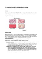
The skin is comprised of three main layers, the hypodermis, dermis and the epidermis (See Fig. 1). These three layers co-exist, what affects one will somehow affect the others. The hypodermis is not strictly considered to be part of the skin, but its role is of equal importance as the epidermis and dermis.
The Hypodermis (Subcutaneous Layer)
Lying just underneath the dermis, the subcutaneous layer is the base foundation and the innermost layer of the skin. This is the layer responsible for the production and storage of fat, which is a combination of areolar tissue (elastic fibres which give the skin flexibility and elasticity) and adipose tissue (containing fat cells).
Fat is a poor conductor of heat, therefore the hypodermis also serves to conserve body heat and as a protective cushion for internal organs.
The hypodermis also contains deeper hair follicles, sweat glands and blood vessels.
The subcutaneous layer is thinner in men than women, which is why women are rounder in shape, however as we grow older the fatty tissue within the subcutaneous layer diminishes and elasticity is lost, which is why wrinkles develop. However, this layer can still be as much as three centimetres thick on the abdomen. Cellulite (the dimpled appearance of skin on some areas of the body) comes from the structure of fatty tissue in this area.
The Dermis
The dermis is positioned just under the stratum basale layer of the epidermis. The dermis consists of connective tissues, and a matrix which binds it together comprised of elastic and collagen fibres (responsible for around 95% of the dermis).
The dermis is around 2mm thick and contains the following structures:
Blood vessels
Lymph vessels
Somatic (sensory nerve endings)
Sweat glands and ducts
Hairs,
Arrector pili muscles
Sebaceous glands
Blood vessels
Blood vessels are hollow tubes which are used to transport blood around the body; blood vessels include a network of arteries, capillaries, veins, venules and arterioles.
Lymph vessels
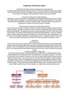
Lymph vessels form part of the system which transports excess protein, water and waste from connective tissues in the body to the blood stream. Whilst the lymph is transported it is cleansed and the lymph vessels are partly responsible for any immune reactions which may take place.
Somatic (sensory nerve endings)
Somatic (sensory nerve endings) are part of the somatic sensory system, which serves two primary purposes, to detect mechanical stimuli (For example touch, pressure, vibration) and to detect painful stimuli or temperature. A superficial burn may cause excruciating pain, but severe burns may destroy the sensory nerves and therefore no pain is felt. This process is one of the most important functions of skin.
Sweat glands and ducts
Sweat glands and ducts work to cool the body down when the somatic system detects excessive heat and play an essential role in thermoregulation.
Arrector pili muscles
Arrector pili muscles are made up of smooth muscle cells. One end of the muscle is attached to the connective tissue along the epidermis and the other to the connective tissue surrounding the hair bulb. When the arrector pili muscle contracts the hair stands on end, the muscles pull on the dermal connective tissue, causing small dimples on the skins surface, this results in ‘goose pimples’.
Contraction of this muscle has a limited insulating effect upon the human body, but for animals covered in fur the contraction process increases the layer of trapped air between the hairs, thus adding to the bodys insulating properties.
Sebaceous glands
Sebaceous glands help maintain healthy skin hair. Most sebaceous glands open into the upper part of a hair follicle, but they can open directly to the top of the skin (e.g. the lips). Sebum is a by product of the sebaceous glands, which can be found on most parts of the body excepting the soles of the feet and the palms, the sebum produced keeps the hair waterproof and supple. However, emotional stress can stimulate the flow of sebum.
The primary role of the dermis is the supply the epidermis with nutrient-saturated blood and to regulate temperature. The dermis also acts to protect the skin from infections which manage to pass through the epidermis, should it fail to pick them up. The dermis is comprised of collagen connective tissues, produced by fibroplasts. Nerve endings, pressure receptors and the root of hair follicles can be found within the dermis.
The Two Layers of the Dermis
The dermis has two distinct layers:
The papillary dermis
The reticular layer
Papillary Dermis
The papillary dermis is the hub of dermis activity, processing the previously mentioned functions of temperature control and the supply of much needed nutrients to the layers of the epidermis which need them. A thin but very extensive vascular system, similar to that on other parts of the body is used to achieve these functions. Blood flow through the skin is controlled by expansion and restriction; this also determines how quickly heat is dispelled when a temperature change occurs.
The Reticular Layer
The reticular layer is far denser than the papillary dermis, and this provides the skin with shape, strength and a certain amount of elasticity, which decreases with age. This layer also serves to support other skin components, such as hair follicles, sweat glands and sebaceous glands.
The Epidermis
The epidermis is the outside layer of cells that serve as a protective shield for the body. It consists of a thin layer of closely packed cells approximately 0.12mm thick. It is considerably thicker in areas subjected to constant pressure or friction such as the soles of the feet or the palms of the hand. The cells of the epidermis only live for about one month (around 14 days for infants, rising to 36 days for the over 50s), therefore, the epidermis is constantly regenerating itself.
The epidermis consists of five layers as follows:
Stratum Germinativum / basal layer (bottom layer)
The basale layer as it is also known, is situated directly above the dermis and is the deepest layer of the epidermis. Here, the process of mitosis (cell regeneration) commences. This layer is also known to be responsible for the production of melanin, thus protecting us from ultraviolet rays. The stratum corneum (see below) relays messages to this layer, when frequent pressure is applied on a particular part of skin (such as the soles of the feet) thus stimulating the creation of more skin cells, which results in a thicker layer of skin growing.
Melanin Production
Melanocytes are a single layer of cells which occur in the deepest layer of the epidermis, from which melanin is produced. This pigment is transferred to hair cells and the epidermis and gives hair and skin its colour. Another function of melanin is to provide protection from ultraviolet rays, when exposed to ultraviolet light the skin darkens, providing the body with yet more protection as needed. People living in hot climates often have higher quantities of melanin in their body.
Keratinocyte cells ingest melanin granules, there they gather around the nucleus of the keranocite. Melanin is a substance which absorbs the ultraviolet light before it can reach the nucleus; melanin protects the DNA in the nucleus from UV radiation damage. Keratin is a protein found only in the hair, skin and nails. It consists of primarily dead skin cells, which themselves are almost entirely made of protein.
Stratum Spinosum / prickle cell layer (Fourth layer)
This is where the process of keratinisation commences, nourishment from the protoplasm (tissue fluid) is received through filaments (threads which connect the tissues), already the cells are starting to flatten in shape at the top. This is a slightly thicker layer and contains 3-6 layers of cells. Desmosomes are keratin based bridges which form in between the cells which allow them to move more freely between one another. Division of the cells in this region is ongoing.
Stratum Granulosum / granular layer (third layer)
The stratum granulosum is so called because the cells are granular (polygonal) in shape. This layer is around 1-3 cells in thickness. As the cells grow, the nucleus disintegrates and dies. The cells themselves flatten as fluid which would have allowed them to turn into keratin is lost, this is the final stage of keratinisation. The outermost cells in this layer are dead.
Stratum Lucidum / transparent layer (second layer)
Cells in the stratum lucidum are tightly packed, transparent and contain no nucleus. It is believed that this is a layer which acts as a barrier to the transmission of water. In some areas of the body this layer is quite thick yet absent in other areas entirely. This layer is also responsible for the root of nail growth.
Stratum Corneum / horny layer (the outer layer)
The stratum corneum is the outermost layer of the skin and consists of dead skin cells, ready to be shed as a result of abrasion, for example friction with clothing or washing of the face. This layer is very thin, often only 5-15 microns thick. As the stratum corneum sheds cells, others from the stratum lucidum move outwards to replace the old lost cells.
The cells underneath this level contain an epidermal fatty substance which makes the skin waterproof and prevents the skin from cracking; this substance also acts to prevent bacteria from being unduly absorbed.
Changes to the epidermis
Age
Ageing of the skin is a natural process. As we grow older our skin produces less sebum and changes occur in the collagen elastin fibres. The skin becomes thinner as the rate of mitosis slows down. Cell regeneration decreases and we loose muscle tone causing sagging on the cheeks. Dilated capillaries can often appear around the nose and cheeks. Liver spots may appear around the temples and on the hands.
Sunlight
The suns ultraviolet radiation can penetrate into the dermis and causes dehydration and also affects the collagen and elastin fibres. This can cause the skin to loose its strength resulting in wrinkles and sagging.
Medication
Certain medications can lead to skin dehydration, fluid retention, swelling, sensitivity and allergies. Antibiotics can cause dryness of the skin, steroids can swell the tissue and the contraceptive pill can cause chloasma which results in pigmentation occurring usually on the face. Creams such as hydrocortisone can cause thinning of the skin if used for long periods of time.
Environment
Air pollution such as carbon from smoke fumes from car exhausts, chemicals from factories and the environment, can all be potentially damaging to the skin's cells and can dehydrate the skin. Environmental pollutants such as lead, mercury, aluminium can all accumulate in the body, and may find their way into food through polluted waters, rain and dust. Central heating can lead to a dry environment which affects the moisture content of the epidermis.
Extremes of temperature can damage the skin. A cold climate can lead to a reduced production of sebum, resulting in reduced protection for the skin and increased water evaporation.
A hot climate can lead to increased moisture loss through perspiration and can cause the capillaries to constantly contract and dilate which leads to the walls weakening causing permanently dilated capillaries (thread veins). This is common on the cheeks and nose.
Chemicals
Daily exposure to household irritants, chemicals, DIY materials, regular washings with hot water and soap, can remove natural oils from the surface of the skin leaving its deeper layers unprotected. With the natural oil stripped from the outer layer of skin, its natural moisture (water) quickly evaporates from the surface of the skin, turning it dry, irritated, and itchy. Dry cracked skin easily gets infected, the inflammation of the skin can, in its turn, become a base for such serious skin diseases as psoriasis and eczema. Acids and Alkalis can cause skin burns and chemical detergents may lead to allergic reactions, sensitivity, or an exacerbation of skin conditions including dermatitis, eczema, and inflammation.
Other factors include;
Alcohol - Alcohol has dehydrating effect on the skin, and excess consumption causes blood vessels in the skin to dilate.
Smoking - Affects the skin's cells, and destroys vitamins B and C which are important for a healthy skin. Smoking dulls the skin by polluting the pores and increases the formation of lines around the eyes and the mouth.
Diet - A healthy diet is needed for healthy skin, a nutritionally balanced diet is vital to the skin, as it will contain all the essential nutrients to nourish the cells in growth and repair. In addition to the essential nutrients in the diet, the following vitamins aid the regeneration of the skin: Vitamin A helps repair the body's tissues and helps prevent dryness and ageing. Vitamin B helps improve the circulation and the skin's colour, and is essential for cellular oxidation. Vitamin C is essential for healing and to maintain levels of collagen in the skin.
Water - Drinking an adequate amount of water aids the digestive system and helps to prevent a build up of toxicity in the tissues of the skin.
Exercise - Regular exercise promotes good circulation, increased oxygen intake and blood flow to the skin.
Sleep - is essential to physical and emotional well being and is one of the most effective regenerators for the skin.
Summary
The skin is a very intricate mechanism which performs many functions; the skin is divided into a number of complex layers, which interact with one another to perform the many tasks it must carry out. Actions which affect the skin may have an effect upon another part of the body entirely.
Bibliography
Useful Literature
Anatomy and Physiology, in Health and Illness, Anne Waugh & Allison Grant, Ross & Wilson, 10th edition, 2006
Indian Head Massage, Francesca Gould, Nelson Thornes, 2002
Indian Head Massage, Helen McGuinness, Hodder & Stoughton, 2007
Indian Head Massage, Narenda Mehta, Thorsons, 2001
The New Book of Massage, Lucy Lidell & Sara Thomas, Ebury Press, 2000
Diagram of the skin