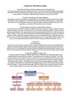
How T Lymphocytes Recognise Antigens: T lymphocytes (Human Biology Book, pages 136-138) are part of the immune surveillance system. They help identify antigens, which are substances foreign to the body. However, to be recognised by a T lymphocyte, an antigen must be processed and "presented" to the lymphocyte in a form it can identify, shown below.
1. An antigen circulating in the body has a structure that a T lymphocyte cannot recognise.
2. An antigen-processing cell, such as a macrophage, engulfs the antigen.
3. Enzymes in the antigen-processing cell break the antigen into fragments.
4. Some antigen fragments become linked with major histocompatibility complex molecules and are then shuttled to the surface of the cell membrane.
5. A T-cell receptor, located on the surface of a T lymphocyte, recognises the antigen fragment linked with a major histocompatibility complex molecule and binds to it.
Mobilisation: Once an antigen has been recognised by an antigen-processing cell and T lymphocyte, a series of events to activate the immune system follows. When an antigen-processing cell ingests an antigen, it releases cytokines, for example, interleukin-1, interleukin-8, or interleukin-12, which act on certain other cells. Interleukin-1 mobilises other T lymphocytes; interleukin-12 stimulates natural killer cells to become more potent killers and to emit interferon; interleukin-8 acts as a sign, guiding neutrophils to the site where the antigen was spotted. This process of attracting and recruiting cells is called chemotaxis.
When T lymphocytes are triggered through their T-cell receptors, they produce a lot of cytokines that help to recruit other lymphocytes, therefore amplifying the immune response. Cytokines can also start the non-specific (innate) immune defences. Cytokines therefore link innate and learned immunity.
Attack: Much of the immune system's machinery is aimed at killing or eliminating invading microbes once they have been recognised. Macrophages, neutrophils, and natural killer cells are able to eliminate many foreign invaders.
If an invader cannot be eliminated completely, walls can be built to imprison it. The prison wall is made of special cells, called a granuloma. Tuberculosis (See Biological Science book pages 456-457) is an example of an infection that is not totally eliminated. The bacteria that cause tuberculosis are imprisoned within a granuloma. Most healthy people who are exposed to these bacteria fend off the tuberculosis infection, but some bacteria survive indefinitely, usually in the lung, surrounded by a granuloma. If the immune system is weakened (even 50 or 60 years later), the prison walls deteriorate and the bacteria that cause tuberculosis start to multiply.
The body doesn’t fight all invaders the same way. Invaders that stay outside the bodies cells (extracellular organisms) are quite easy to fight; the immune system mobilises defences to facilitate their intake by macrophages and other cells. How the immune system does this depends on whether the invaders are encapsulated (have a thick capsule around them) or are non-encapsulated. Invaders that gain access to the inside of cells (intracellular organisms) and remain viable (alive) and functional are fought in a different way altogether.
Before vaccines (See Biological Science book pages 533-539) were invented many people died of illnesses that today are prevented. Some examples of these illnesses are whooping cough, measles, and polio. Those same germs exist today, but vaccines now protect people. There are a series of steps that your body goes through in fighting off a vaccine-preventable disease:
First: A vaccine is given by a shot or liquid by mouth. An alternative needle-free way is the use of inhalation by aerosol and powder. Most vaccines contain a weakened or dead disease germ or part of a disease germ. Other vaccines use inactivated toxins. Some of the bacteria that cause disease do so by producing toxins that invade the bloodstream.
Next: The body makes antibodies against the weakened or dead germs in the vaccine.
Then: These antibodies can fight the real disease germs -- which can be lurking all around -- if they invade the child's body. The antibodies will know how to destroy them and the child will not become ill. Most vaccines don't cause the diseases that are usually caused by viruses and bacteria.
Finally: Protective antibodies stay on guard in the child’s body to safeguard it from the real disease germs.
Medical Developments
Monoclonal Antibody (MAb). This is a laboratory-produced protein molecule used in medicine to detect pregnancy; diagnose disease, including acquired immuno-deficiency syndrome (AIDS), hepatitis, and various kinds of cancer; and treat conditions caused by toxins (poisonous substances), such as snake venom.
When MAbs were first discovered in the 1970s, scientists expected they would revolutionise the way diseases such as cancer are treated. Over 20 years later, MAbs best serve medicine as analytic tools and treatment aids—that is, that is when combined with more conventional therapies. They are also used in laboratories to track proteins in various experiments.
Formation and Structure: An antibody is a Y-shaped protein produced by a type of white blood cell known as a B cell. B cells are made in the bone marrow of the body in response to a foreign substance (an antigen). Antibodies neutralise or mark antigens for destruction by other cells of the immune system. Antibodies perform their work by attaching—or binding—to specific parts of antigens called receptors. Only antibodies created for a specific antigen can attach to that antigen’s receptors. Once an antibody is produced, it remains in the blood, ready to attack its targeted antigen the next time the antigen invades the body. As a result, the blood contains hundreds of thousands of different types of antibodies. These antibodies mix freely, making it difficult to isolate a particular type.
How Monoclonal Antibodies Are Made: A monoclonal antibody is created in the laboratory by joining together a normal B cell and a B cell affected with multiple myeloma, a type of cancer that causes a single B cell to reproduce in large quantities. This fusion creates a hybrid cell, or hybridoma, that produces an unlimited supply of the antibody secreted by the original normal B cell. By varying the types of normal B cells used to create hybridomas, scientists can create many different kinds of MAbs.
Immunologists Georges J. F. Köhler and César Milstein created the first MAbs in 1975. Scientists had earlier discovered that mouse myeloma cells produced large quantities of an antibody for a specific antigen. However, the antibody they secreted had no medical use. Köhler and Milstein developed a technique that combined the cancerous B cell’s ability to rapidly produce large quantities of the same antibody, with the ability of a normal B cell to produce a useful antibody. The two researchers grew normal mouse B cells and mouse myeloma cells together in a laboratory culture. The growing medium included a chemical that would join together the membrane of one normal B cell with the membrane of one myeloma cell, creating a B cell hybridoma. Before the cells were fused together, however, the deoxyribonucleic acid (DNA)—the chemical that carries the genetic material of an organism—was removed from the myeloma mouse cell. The DNA for the normal B cell then became the DNA for the new hybrid cell. Köhler and Milstein separated each hybridoma from the culture and placed it in its own growing medium. Each cell grew and multiplied at the rate of the original mouse myeloma cell from which it was derived, but all of the new cells produced secreted only the antibody made by the original normal B cell used to create the hybridoma. Köhler and Milstein received the Nobel Prize for Physiology or Medicine in 1984 in recognition of this work.
Current and Future Research: In the past ten years, researchers have begun to develop more sophisticated techniques for generating MAbs. Human B cells can now be grown in mice to which human cells or human antibody genes (the hereditary units that determine the particular characteristics of an organism) have been transferred to create a human immune system. These B cells are used to create MAbs that are mostly human in composition. By disguising a mouse antibody as human, researchers hope to fool the immune system of a human patient into accepting the antibody as one of its own. Also, the cloning of the genes of antibodies now allows scientists to rearrange these genes to produce MAbs that are smaller and more effective in penetrating a solid tumour. Recently, the field of bispecific MAbs has appeared. This technology allows scientists to fuse two hybridoma cells together to generate hybrid-hybridomas that give out MAbs with two different sets of binding sites (the areas where they attach to antigen receptors). For example, one binding site may recognise a tumour cell, and the other site may recognise a cell or toxin that can be recruited to kill the tumour cell. These bispecific MAbs can also be produced bio-chemically by using chemicals to join individual proteins or genetically by linking the genes for the different MAbs. In cancer therapeutics, a lot of interest is being focused on designing MAbs specific not only for molecules on tumour cells but also for molecules produced on actively growing blood vessels. Solid tumours require a constant supply of blood to survive. One way they ensure this supply is by encouraging the growth of new blood vessels that produce unique growth-related molecules. Normal blood vessels do not grow actively and do not produce this molecule. By injecting a patient with the new type of MAb, scientists hope to destroy only the blood vessels associated with a tumour, depriving it of nutrients and eventually killing it. In the near future, modern advances in the design and production of MAbs will result in the creation of more efficient versions of these special antibodies. At that point, practitioners will be able to choose from a substantial collection of clinically effective MAbs that they can use to treat their patients.
Bibliography
I gathered all of the information that I used from:
-
The National Immunisation Program (NIP) web site. I accessed this site via .
2) The Merck Manual of Medical Information--Home Edition. I gained access to this via .
3) A New Introduction to Human Biology AS by Bill Indge, Martin Rowland and Margaret Baker (Hodder & Stoughton 2000.
4) Human Biology book by Mike Boyle, Bill Indge and Kathtyn Senior (Collins 1999).
5) Biological Science 1 & 2, Cambridge, N.P.O. Green, G.W. Stout, D.J. Taylor, and the editor R. Soper. 1984, and 1990.