The Structure and biological functions of cell membranes
Andrea McCaffery
HEFC Biological Science
The Structure and biological functions of cell membranes
Cells are the fundamental units of life, because a cell is the simplest unit capable of independent existence.
Biological membranes maintain the spatial organisation of life.
Cell membranes define the boundaries of living cells and work to shield it from changes in its environment. Essentially, membranes prevent undesirable agents from entering cells and keep needed molecules on the inside. Therefore, the cell membrane controls and regulates everything that passes in or out of the cell.
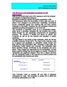
The membrane is essentially made up of a phospholipid bilayer, which forms a boundary enclosing the cell contents and is also folded through the cell, separating compartments for specialised purposes. The structure is flexible and allows for growth and movement as well as for the insertion and operation of protein machinery.
The lipid bilayer is two layers of lipid molecules, which are termed amphililic (meaning "loving both"). The head of the molecule, made up of glycerol and phosphate, is hydrophilic ("water loving") and polar and the long hydrocarbon tails, composed of fatty acids, are hydrophobic ("water hating") and non-polar. The hydrophilic head bears electrical charges contributed by the phosphate and some bases. These charges are responsible for the hydrophilicity. No lipid bears a positive charge they are all negative or neutral. Therefore all membranes are negatively charged. They form a bilayer structure with the hydrophilic heads to the external and internal surface and the hydrophobic tails towards each other.
Lipid molecules make up between 30 and 80% of biological membranes by mass. The remainder is protein (20 to 60%) and sometimes carbohydrate (0 to 10%).
Within the lipid structure there is also a certain amount of cholesterol embedded between the phospholipids. This serves to increase the strength of the membrane and makes it a little less fluid.
Andrea McCaffery
HEFC Biological Science
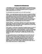
The protein molecules are very much larger than the lipid molecules. Proteins are responsible for moving molecules and messages across the membrane and they are an integral part of the membrane itself. They are not fixed rigidly and can diffuse across the surfaces of cells relatively easily and independently and within the plane of the membrane. Proteins appear in many different forms. The Intrinsic or transmembrane protein spans the width of the membrane with its hydrophilic parts at either surface and its hydrophobic parts sandwiched between the hydrophobic tails of the lipids. These provide a control channel through the membrane to allow small molecules such as water a passage in or out of the cell. Extrinsic proteins are associated with the membrane surface. These proteins act as receptors to stimuli e.g. hormones. There are also Glycoproteins (proteins and carbohydrates) and Glycolipids, which are positioned on the outer surface only which act as recognition markers for things such as tissue types and blood groups etc.
The function of a cell membrane, as well as being to separate the cell from its external environment and act as a receptor site, is to be a surface upon which certain metabolic pathways can occur. This semi permeable barrier controls various mechanisms of transport.
The most simple method of transport, which takes place across a plasma membrane, is diffusion. This mechanism involves small, uncharged solutes moving easily from a high concentration to a low concentration.
Similarly, the process of Osmosis occurs when water molecules move down their concentration gradient across a partially permeable ...
This is a preview of the whole essay
The function of a cell membrane, as well as being to separate the cell from its external environment and act as a receptor site, is to be a surface upon which certain metabolic pathways can occur. This semi permeable barrier controls various mechanisms of transport.
The most simple method of transport, which takes place across a plasma membrane, is diffusion. This mechanism involves small, uncharged solutes moving easily from a high concentration to a low concentration.
Similarly, the process of Osmosis occurs when water molecules move down their concentration gradient across a partially permeable membrane.
It is most important in animal cells that the movement of water in or out of the cell is kept to a minimum. The effect of too much water moving into the cell would cause the cell to expand and lyse. As a results of too much water leaving the cell it would collapse inwardly
Andrea McCaffery
HEFC Biological Science
and the concentration of the cytoplasm would lose its internal structure and cease to function. This is called plasmolysis.
In the case of plant cells the situation is different because of the presence of the cellulose cell wall. Although it is freely permeable, it exerts an inward pressure on the cell so that water cannot enter the cell by osmosis indefinitely and, once full, will remain at its desirable state.
Another mechanism of transport is facilitated diffusion, which allows large molecules to cross the membrane down their concentration gradient. An example of this occurs in liver cells, where rotating proteins carry glucose and amino acids down their concentration gradient with no energy required.
Active transport is a mechanism which carries molecules up the concentration gradient. This requires energy to be used in the hydrolysis of ATP.
H2O
ATP --> ADP + phosphate + chemical energy
It requires a pump protein to transport the substance against its concentration gradient. This mechanism is used, for example, in transporting sodium out of red blood cells.
There is another, very different, form of transport that takes place within the cell membrane and that is bulk transport, where larger particles need to enter or leave the cell. Membrane transport systems cannot do this type of job, but the cell membrane itself can.
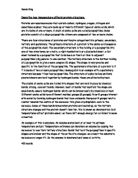
Endocytosis is the term used when particles are surrounded by and taken up into the membrane-lined vesicles. This can occur at a relatively large scale, for example, when bacteria are ingested.
In this case the process is called phagocytosis -"cell eating" and the alternative process is pinocytosis -"cell drinking". Cells that specialise in this are refered to as phagocytes and a white blood cell is a good example of this. See diagram.
Andrea McCaffery
HEFC Biological Science
Exocytosis is the opposite process which involves the secretion of material through the cell membrane.
The Golgi apparratus is a system of tubular structures formed by plasma membrane. The Golgi receives vesicles from the endoplasmic reticulum. These vesicles containing material such as proteins and hormones then divide way from the Golgi body, fuse with the plasma membrane and are exported from the cell where the molecules release into the tissue fluid.
For transport reasons, some specialised membranes have different structures according to their functions.
The nucleus, which controls the activity of the cell, has a double membrane but this is perforated by many nuclear pores to allow the movement of RNA. The relatively small messenger RNA molecules need to be able to pass easily through the pores in the nuclear membrane, carrying genetic messages from the nucleus to the cytoplasm. They then move to the surface of the ribosomes, transporting the instructions to the site of protein sythesis.
The Mitochondria is also very specialised. It is said to be the cellular power station and has many important functions.
It is the only site of cellular respiration and electronic transport.
Through a process known as the "Kreb cycle", the matrix of the Mitochondria containing enzymes to carry out the reactions of respiration, converts carbohydrates to carbon dioxide.
In addition, along the inner membrane folds (cristae) there is an electron transport chain which adds protons to convert O² to H²O to make ATP energy. Cells which require a lot of energy will have many Mitochondria. They also contain their own gentic material, so that when a cell divides, the Mitochondria replicate. Mitochondria also replicate at times other than cell division, for example when the long term energy demands of a cell increase.
Andrea McCaffery
HEFC Biological Science
The Mitochondria's specialised membrane structure is perfectly adapted to carry out their important functions.
The Mitochondria has a double membrane. The outer membrane allows small molecules such as glucose to pass through freely but larger molecules are excluded. The inner membrane has many folds. These give a much increased surface area on which the vital chemical reactions can occur.
Chloroplasts in plant cells have a structure that is in some ways similar to that of the Mitochondria. Chloroplasts exist only in plant cells and not in animal cells. The pressence of chloroplasts is most important because they enable plants to make their own food.
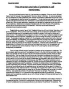
Throught the process of photosynthesis their function is to use light energy to convert carbon dioxide and water into glucose and oxygen.The equation for this is shown below.
6CO2 + 6H2O -------> C6 H12 O6 + 6O2
Like Mitochondria, they contain their own DNA and have a double membrane. Similarly, chloroplasts have an enormously folded inner membrane which gives a greatly increased surface area on which biological reactions take place.
This is another example where the specialised structure of the membrane is to provide the optimum environment in which to perform its vital functions.
To conclude, we can see how complex and how infinately paramount the structure and functions of a biological cell membrane are to all living things.
Membranes are of crucial importance to life, because a cell must separate itself from the outside environment for two major reasons. It must keep its molecules of life (DNA, RNA, proteins) from dissipating away and it must keep out foreign molecules that damage or destroy the cells components and molecules. However, while the cell must always abide by these two principles, it must also communicate with the environment to continuously monitor the external conditions and adapt to them.
Biological membranes are bilipid layers. The bilayer has a head which is the negatively charged phosphate group and two tails which are two highly hydrophobic hydrocarbon chains of the phospholipid. The tails of the phospholipids orient towards each other forming a hydrophobic environment within the membrane and the charged phosphate groups face out into the hydrophilic environment.
Andrea McCaffery
HEFC Biological Science
This semi-permeable bilipid layer allows some smaller molecules to diffuse freely through the membrane.
To carry out the more complex transport systems of moving larger molecules across the membrane, the membrane has a large number of proteins embedded in it.
These can pass all the way through the membrane (Intrinsic proteins), providing a passage for molecules, or can rotate between the surfaces (Extrinsic proteins), acting as receptors to such things as hormones and to act as recognition markers.
As well as diffussion, we have looked at the other more complex mechanisms of transport such as osmosis, facilitated diffusion and active transport which take place across the cell membrane. The other processes, Endocytosis and Exocytosis, involving the Golgi Apparratus are the methods of transport for inporting and exporting even larger molecules through the cell membrane. Using these methods of transport, cell membranes control the inward and outward movement of useful molecules.
We have also seen that there are a number of specialised membranes with slightly different structures according to there particular function. For example, we have looked at the way in which the membranes of the Mitochondria and the chloroplast have adapted to best suit there function and how the two have a similar double membrane with multiple folds in the inner membrane. Although for the same fundemental purpose ; to increase the surface area, the inner membrane of the Mitochondria and the chloroplast are the sites for completely different chemical reactions.
The structures of biological cell membranes are designed in such a way as to aid their very function. It is the interface between a cell and its environment and a fluid structure which preforms the selective transport of essential materials into and out of the cell.
Andrea McCaffery
HEFC Biological Science
References
S.J. Singer and G.L. Nicholson (1972) The fluid mosaic model structure of cell membranes.
Fundemental Principles of Membrane Biophysics - David Njus, Dept of Biological Sciences, Wayne State University
Heineman Advanced Science - Biology, Ann Fullick.
4