Membranes are extremely small and thin, so to examine them you need to use an electron microscope. Under the electron microscope they look like solid structures, but they actually move constantly, joining with other membranes. By using the electron microscope we can deduce that the membrane has three layers, two dark layers which are protein with a light layer in the middle which is the bilayer. The total thickness of the membrane is 7nm.
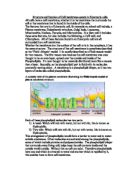
In 1972 Singer and Nicholosen came up with their theory abut the structure and function of the cell membrane. Their theory is known as the Fluid Mosaic Model. Whether the membrane is at the cell surface or in its cytoplasm it has the same basic structure. Structurally, the membrane is a lipid bilayer, which generally means it has two layers. The layers are composed of a two part molecule called a phospholipid. The phospholipid bilayer is composed of various cholestrol, phospholipids, glycolipids and proteins. There are not many strong chemical bonds holding membranes together. Instead membranes get their strength, great flexibility and mobility by a large number of weak interactions resulting from the properties of lipids.
Permability studies showed that lipids could move easily between the interior of cells therefore it was decided that the cell membrane must contain lipids to allow fat soluble materials such as glucose, amino acids and ions to move across the membrane by being transported in it. In membranes the lipids are of a special kind called amphipathic lipids. The second type is the lipids bilayer or a `micelle’ which is a single layer circle of phospholipids with the fatty acyl tails pointing inwards.
The basic structural unit of virtually all biomembranes is the phospholipid bilayer. The bilayer is a sheet like structure composed of two layers of phospholipid molecules whose polar head groups face the surrounding water and whose fatty acyl chains form a continuous hydrophobic interior. Each phospholipid layer is called a leaflet. These two layers of phospholipids are made up of hydrophobic (tails) and hydrophilic (heads) molecules.
The lipids or fatty acyl tails are `water fearing’ (hydrophobic) molecules. The phosphate end is water loving (hydrophilic). The membrane forms when the phosphate head faces out, attracted to the watery environment of the cell and the lipid faces inwards trying to avoid the water.
The hydrophilic heads are polar, so they are bery soluble in water and when they point outwards they form hydrogen bonds with water. The hydrophobic tails are non-polar, so they are very insoluble in water and when they point towards one another it maximizes hydrophobic attractions and excludes water. Due to the different solubility properties of the two ends of phospholipid molecules a phospholipid bilayer can act as a barrier between two aqueous environments.
Phospholipids are two fatty acids, one saturated and one unsaturated which are linked to a glycerol. Differences in lipid composition affect the fluidity and permability of the membrane. The unsaturated fatty acid tails are `kinked’ created by a double carbon bond, which limit close packing of the hydrophobic tails and so increase fluidity, but cholestrol may interfere with the lateral movement of the hydrophobic tails causing a reduction in membrane fluidity.
The smaller molecules between the phospholipids is cholestrol. Cholesterol and its derivatives constitute another importat class of membrane lipids, the steroids. Cholesterol is the major constiuent of animal tissues. Although cholesterol is almost entirely hydrocarbon in composition it is amphipathic because it contains a hydroxyl group that interacts with water. Cholesterol in the membrane interacts with adjacent phospholipids making the membrane more rigid or stable.
Another impritant but minor constituent of membranes is carbohydrate. When carbohydrate is attached to proteins it forms glycoprotein and when it is attached to lipids it forms glyclipid. In the plasma membrane, all the O- and N- lined oligosacharides in glycoproteins and all of the oligosacharides in glycolipids are on the exoplasmic surface. In the endoplasmic reticulum they are found on the interior membrane surface. Surface carbohydrates are known collectibely as glycocalyx and are usually oligosacharides which are positioned to aid in cell recognition functions. Glycocalyx act as antigens and are important in immune response.