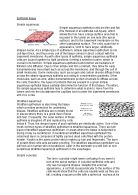
Muscle tissue consists of fibres (cells) that are constructed to generate force. As a result of this characteristic, the function of muscle tissue is to produce motion, maintain posture, and generate heat. Based on its location and certain structural and functional characteristics, muscle tissue is classified into three types: skeletal, cardiac, and smooth (Tortora and Garbowski, 2000). For example, muscle tissue (figure 1.1 C) consists of long, slender, muscle fibres. These muscle fibres slide over one another thereby creating movements (Layman, 2003).
Connective tissue is the most abundant and widely distributed tissue in the body (Tortora and Garbowski, 2000). Connective tissue (figure 1.1 B), in great contrast, includes a lot of intercellular material between its cells. Frequently, this intercellular material contains long, slender rods – connective tissue fibres. Such fibres help connective tissue do its main job, which is to directly or indirectly connect body parts together (Layman, 2003).
Nervous tissue forms a network of nerve cells in the body and also in the brain and spinal cord (Hall, 2005). Also it is the major tissue for communication and control within the body’s internal environment. The nervous tissue (figure 1.1 D) largely does its communicating by means of neurons, i.e. the nerve cells. For example, a function of neurons within the nervous tissue is to inform the brain when the body has been damaged, usually resulting in the sensation of pain (Layman, 2003).
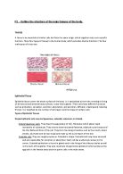
Diagram 1 is Pseudostartified columnar ciliated epithelial tissue. Diagram 2 is Compact bone (connective) tissue. In diagram 1 is a section view of pseudostratified columnar epithelium of trachea. In diagram 2 is sectional view of an osteon (Harvesian system) of femur (thigh bone).
Diagram 1
Diagram 2
Pseudostratified columnar ciliated epithelium
As the name implies, pseudostratified epithelium (diagram 1) is not a true stratified tissue. Cells are columnar but tall and thin. All cells are attached to basement membrane, but not all reach the apical surface (Tortora and Garbowski, 2000). From this can be noted, that the unique appearance of pseudostratified epithelia occurs because of the tall, thin cell intertwine. Nuclei appear at various levels but there is no distinct layering. The elongated shape is what causes the nuclei not to line up and add to the pseudostratified appearance. Columnar epithelium can be compressed a great deal, allowing the tissue to change shape. For example, compressed epithelium is important for the respiratory tract, especially the bronchi, so it can expand and contrast as human breathes in and out (Epithelial cells, 2010). In addition this tissue is most prevalent in the upper or lower respiratory tract, for example trachea. Its function is to line the cavities of the body involved in respiration, serving as a barrier between air coming into the respiratory system and their inner tissues of the body. It also warms and cleans the air before it reaches the lungs, where oxygen from the air is absorbed into the bloodstream (Tissues of the human body, 1999). For example, cilia beat in a rhythmic manner to propel mucous in order to carry unwanted particles along with mucous out of the system to maintain its cleanliness (McGuiness, 2002).
Compact bone
According to Clancy and McVicar (1995), Bone is a tough and rigid tissue (diagram 2). The mature cells are called osteocytes, and the protein fibres present are similar to collagen, which is also present in the bone. The matrix is made up of inorganic salts of calcium and these give bone its hardness. Compact bone tissue is arranged in units called osteons or Haversian systems, which makes up concentric circles called lamellae. Each Haversian canal contains an artery, a vein, lymph vessels and nerve that run through it. Bone is a very dynamic tissue, constantly being restructured as older osteons are replaced by new ones. Between the lamellae (lamellae fibres gives strength to the compact bone) there are small spaces called lacunae, which contains osteocytes – bone cells. Radiating in all directions from the lacunae there are tiny caniliculi, which are filled with extracellular fluid. The caniculi connect lacunae with one another and with the central canal. For example, blood-borne nutrients and oxygen can diffuse to and from the bone cells from the central canal through these canaliculi. Osteons in compact bone tissue are aligned in the same direction along lines of stress. In the shaft, for example, they are parallel to the long axis of the bone. As a result, the shaft of a long bone resists bending or fracturing even when considerable force is applied from either end. The lines of stress in the bone are not static. For example, when person undertakes weight training, lines of stress change in response to repeated strenuous physical activity. Thus, the organisation of osteons changes over time in response to the physical demands placed on the skeleton. Compact bone tissue provides protection and support. For example resists the stresses produced by weight and movement (Tortora and Garbowski, 2000).
The aforementioned evidence shows that human body is compiled of different cells that work together in a group with specialised functions, which forms different kinds of tissues, for example, epithelial tissue that lines the cavities and surfaces of structures throughout the body, furthermore, different tissues form organs.
References
Boyle, M. and Senior, K. (2008), Human Biology, 3rd edition, London, Harper Collins Publishers Ltd
Clancy, J. and McVicar, A. (1995), Physiology and Anatomy, London, Hodder Headline Group
Gersh, I. (2008), Epithelium. Access Science, McGraw-Hill Companies (accessed 20 10 2013)
Hall, D. (2005), The Human Body, Kent, Grange Books Plc
Introduction to Biosolid Mechanics
(accessed 29 10 2013)
Layman, D. (2003), Biology Demystified A Self – Teaching Guide, United States of America, McGraw-Hill Companies Inc
McGuiness, H. (2002), Anatomy and Physiology Therapy Basics, 2nd edition, Bristol, Hodder and Stoughton, Arrowsmiths Ltd
Mallery, C. Animal structure and function (accessed 25 10 2013)
Tortora, G. and Garbowski, S. (2000), Principles of Anatomy and Physiology, 9th edition, United States of America, John Wiley & Sons Inc
‘’Epithelium’’, McGraw-Hill, Concise Encyclopedia of Scinece and Technology, 5th edition, New York, McGraw-Hill Professional, 2005.839 (accessed 20 10 2013)
(accessed 28 10 2013)
(accessed 28 10 2013)
Bibliography
Boyle, M. and Senior, K. (2008), Human Biology, 3rd edition, London, Harper Collins Publishers Ltd (p. 25)
Clancy, J. and McVicar, A. (1995), Physiology and Anatomy, London, Hodder Headline Group (p. 66, 67, 74, 79)
Gersh, I. (2008), Epithelium. Access Science, McGraw-Hill Companies (accessed 20 10 2013)
Hall, D. (2005), The Human Body, Kent, Grange Books Plc (p. 8, 9)
Introduction to Biosolid Mechanics
(accessed 29 10 2013)
Layman, D. (2003), Biology Demystified A Self – Teaching Guide, United States of America, McGraw-Hill Companies Inc (p. 23,24)
McGuiness, H. (2002), Anatomy and Physiology Therapy Basics, 2nd edition, Bristol, Hodder and Stoughton, Arrowsmiths Ltd (p. 10, 12, 17)
Mallery, C. Animal structure and function (accessed 25 10 2013)
Tortora, G. and Garbowski, S. (2000), Principles of Anatomy and Physiology, 9th edition, United States of America, John Wiley & Sons Inc (p.115, 118, 130, 162,163)
‘’Epithelium’’, McGraw-Hill, Concise Encyclopedia of Scinece and Technology, 5th edition, New York, McGraw-Hill Professional, 2005.839 (accessed 20 10 2013)
(accessed 28 10 2013)
(accessed 28 10 2013)