Connective tissues
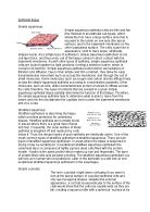
The blood:
Blood is a combination of plasma which is a watery liquid and it is also has cells that float about in it which is why it is called a connective tissue. It is a specific bodily fluid that supplies essential nutrients and substances for our body such as oxygen, sugar and hormones to our cells, and carries waste away from those cells. This waste is finally flushed out of the body in urine, sweat and the lungs. Blood also contains clotting agents; this is caused by platelets clumping together when we bleed to form a blood clot. If exposed to air the platelets break down and release fibrinogen to the bloodstream, this sets off a series of reactions which results in clotting blood on a skin wound which forms a scab. They are two different kinds of blood cells (red and white blood cells). Red blood cells are like slightly indented flattened disks. They’re the most abundant cells, and contain hemogloblin. Whereas white blood cells are cells of our immune system and their purpose is to protect the body against infections and foreign materials.
Cartilage:
Cartilage is a very smooth substance which is found in many areas in the bodies of humans and other animals which include the joints between the bones, the ear, the noes, the elbow, the rib case, the knee, the ankle, the bronchial tubes and intervertebral disks. It is not as hard as bone but it is stiffer and less flexible than muscle. Cartilage is made up of specialised cells called chondrocytes that produce a large amount of extracellular matrix composed of collagen fibres, abundant ground substances rich in proteoglycan, and elastin fibres. They’re three different types of cartilage known as elastic cartilage, fibrocartilage and hyaline cartilage, which differ in the relative amounts of these three main components. Unlike other connective tissues, cartilage does not contain any blood vessels. The chondrocytes supplied by diffusion, helped by the pumping action generated by compression of the articular cartilage or flexion of the elastic cartilage which grows and repairs more slowly.
Areolar tissue:
Areolar connective tissue has no obvious structure, like layers or rows of cells. You might think that this would make it harder to identify. But if you realize that the lack of pattern is one of the distinguishing characteristics of areolar connective tissue, you have learned a cue that will allow you to recognise it. Areolar connective tissue is made of cells and extracellular matrix "extra" means "outside" so the extracellular matrix is material that is outside of the cells. The matrix has two components, fibres and ground substance. In the images on this page, you can see the fibres very easily which look like threads. The only part of the cells that is visible is the nucleus. The ground substance has no structure, so you can't tell that it is there. The ground substance fills all of the spaces between the cells and fibres.
Muscle tissue
Stratified muscle tissue:
The stratified muscle found in the skeletal has an appearance of light sand dark stripes going though it visible using a light telescope. A single skeletal muscle cell is long and approximately cylindrical in shape, with many nuclei located at the edges of the cell.
The muscles are fibrous, dense tissues, which their function is to allow the body to move by repeated contraction and relaxation. Besides movement, the muscle is also responsible for maintaining posture, stabilising the joints, and producing body heat through muscle function.
The movement of striated muscle is controlled voluntarily, unlike the smooth muscle of the internal organs and the cardiac muscle of the heart, which are involuntary. The skeletal muscles are composed of muscle fibres, long fused cells containing multiple nuclei. The muscle fibres are packed together in bundles by connective tissue and are packed with myofibrils. Myofibrils are cylindrical arrangements of myofilaments, made up of proteins called actin and myosin, and which cause the light and dark banded appearance of muscle fibres. These myosin and actin filaments slide across each other, causing the muscle to contract.
Non-striated muscle tissue:
Unlike striated muscle tissue, non-striated tissue is smooth muscle fibres which are small and narrowing with the ends reducing in size, In contrast to the cylindrical shape of skeletal muscle. Each smooth muscle fibre has single centrally locate nucleus.
Contractions of the non-striated muscle constrict which is reduced diameter of the vessels they surround. This is very important in the digestive system in which the action the smooth muscle helps to move the food along the gastrointestinal system as well as breaking the food down further. Smooth muscle also contributes to moving fluids though the body and to the elimination of ingestible matter from the gastrointestinal system.
Cardiac muscle tissue:
Cardiac muscle fibres are striated, branched and sometimes described as Y-shaped. Cardiac muscles are a single central nucleus. These fibres are attached at their ends to adjoining fibres by thick plasma membrane called intercalated disks.
Involuntary muscle only found in the heart regulates the beat at which the heart pumps blood to the arteries which then blood follows to the capillaries and is oxygenated ten follows the veins back to heart and the cycle begins again but before i get off track the main function of cardiac muscle tissue is to regulate the beat of your heart to (in the average human) about 70 beats per minute without getting tired.
Nervous tissue:
A motor neuron a main part of the nervous tissue and has many processes (cytoplasmic extensions), called dendtrites, which enter a large, grey cell body at one end. A single process, the axon, leaves at the other end, extending towards the dendrites of the next neuron or to form a motor endplate in a muscle. Dendrites are usually short and divided while the axons are very long and does not branched freely. The impulses are transmitted through the motor neuron in one direction, i.e. into the cell body by the dendrites and away from the cell body by the axon . The cell body is enclosed by a cell (plasma) membrane and has a central nucleus. Granules, called Nissl, bodies are found in the cytoplasm of the cell body. Within the cell body, extremely fine neurofibrils extend from the dendrites into the axon. The axon is surrounded by the myelin sheath, which forms a whitish, non-cellular, fatty layer around the axon. Outside the myelin sheath is a cellular layer called the neurilemma or sheath of Schwann cells. The myelin sheath together with the neurilemma is also known as the medullary sheath. This medullary sheath is interrupted at intervals by the nodes of Ranvier.
Describe the way these tissues function in two different names organs in the body.
Stomach:
The stomach is an organ of the digestive system. It is an expanded section of the digestive tube between the esophagus and small intestine. Its characteristic shape is well known. The right side of the stomach is called the greater curvature and the left the lesser curvature. The most distal and narrow section of the stomach is termed the pylorus - as food is liquefied in the stomach it passes through the pyloric canal into the small intestine.
- The wall of the stomach is structurally similar to other parts of the digestive tube, with the exception that the stomach has an extra oblique layer of smooth muscle inside the circular layer, which aids in performance of complex grinding motions. In the empty state, the stomach is contracted and its mucosa and submucosa are thrown up into distinct folds called rugae; when distended with food, the rugae are "ironed out" and flat.
-
The next layer is the inner lining of columnar epithelium with goblet and it works in the stomach and digestive tract provides an impermeable barrier against any bacteria that could be ingested but is permeable to any necessary ions.
- The stomach also contains of the nervous muscle which receives stimuli from both external and internal sources which means it is an essential because it tells other parts of the body what is going on.
- The last main tissue of the stomach is the connective tissue like blood which keeps all of the different parts in place acting like glue doing this by its elastic fibres.
Heart:
The heart is the organ that supplies blood and oxygen to all parts of the body. It is about the size of a clenched fist, weighs about 10.5 ounces and is shaped like a cone. Blood is pumped away from the heart through arteries and returns to the heart through veins. The major artery of the body is the aorta and the major veins of the body are the vena cavae.
- In order for the organs to stay alive and function properly, they need nutrients and oxygen. The nutrients and oxygen reach our organs through blood. Blood is delivered to the organs through arteries.Metabolic wastes and Carbon Dioxide occur as a result of the consumption of these nutrients and oxygen by the cells. Again these materials carried by blood, this time by the veins, go back to the heart to collect and clean the blood.
- The heart is also made of cardiac muscle. This type of muscle only exists in your heart. Unlike other types of muscle, cardiac muscle never gets tired. It works automatically and constantly without ever pausing to rest. Cardiac muscle contracts to squeeze blood out of your heart, and relaxes to fill your heart with blood.
- The nervous system makes sure the heart is working by sending impulses by neurons to the heart to make it contract which controls the speed of your heart.
-
The epithelial tissue which is the squamous epithelial tissue I very smooth which allows the blood to flow around the heart fluently and smoothly
Bibliography
19/09/12
19/09/12
19/09/12
-19/09/12
21/09/12
21/09/12
21/09/12
25/09/12