Why the Body Needs Energy? Every living cell within the body needs energy which is used to do work around the body or to produce heat
Fundamental of Atomy and Physiology
Unit 5 Assignment 2
Task 1(P4)
Introduction - Why the Body Needs Energy?
Every living cell within the body needs energy which is used to do work around the body or to produce heat or light therefore if you exercise you get hot and to digest food we produce heat because your body is using up energy.
Food is glucose which contains starch that then turns into carbohydrate which then produces fuel for the body.
The energy in the body is also used when breathing, thinking, eliminates waste, maintain blood pressure, regular body temperature and contract muscles.
What is Energy used for?
There are two rules of energy.
The 1st rule is that energy always comes from a source of energy for example,
* The sun is a producer this is because the sun produces nuclear energy.
* The tree is a primary source of energy because the tree leaves photosynthesise using the sun as a nuclear energy to produce glucose.
* The fire wood is a secondary source of energy because it produces chemical energy
* The fire is a tertiary source of energy because it burns fuel and releases energy an example of this would be heat, light and sound.
Reference:
Class notes hand out 12 / 3 / 2008
The 2nd rule of energy is that it can't be created or destroyed but only changed from one form to another form an example of this would be someone eating food glucose energy would be transferred to the muscle which allows the person to run therefore transferring chemical energy into kinetic energy, heat and sound energy.
Reference:
Class notes hand out 12 / 3 / 2008
Forms of Energy
Energy comes in many different forms an example of this would be fire is a source of heat energy, sun is a source of light energy, ear absorbs sound energy, food is a source of chemical energy which is the most common energy because it always comes from some sort of packaging and you always have to unpackaged it to use and abuse it.
Energy is a chemical bond which unites atoms and molecules together; this means the bond has to be broken into the glucose molecules that have to be released to produce a chemical reaction to release the energy within the bonds.
Molecules
Glucose C6 H12 O6
Burier continues in carbohydrates
This happens inside the cell
Reference:
Class notes hand out 12 / 3 / 2008
What is needed For Releasing Energy?
When you burn firewood there is always a chemical reaction when the energy is released it forms heat and light.
Fuel = glucose + oxygen
The Concept of Energy Metabolism
Metabolism is where the chemical reaction takes place within the body organs,
The chemical reaction will always be involved in using up energy or releasing energy from chemicals such as glucose.
Metabolic reaction produces different life processes however in order for us to be alive we need these metabolic reactions to release energy so that we can digest the food an example of this would be
* Repairing to make new cells
* Movement
* Growth
* Reproductions building hormones
* Sensitivity sending nerve impulses
These are all process that requires metabolism within the body organs.
Catabolic and Anabolic Reaction
This is when glucose is transferred into every cell within your body and is broken dawn into energy however the catabolic reaction means releases energy an example of this will be when carbohydrates --> glucose --> cell --> energy --> mitochondria --> oxygen and anabolic reaction means when energy uses up energy this is when the anabolic steroids build up muscles.
Reference:
Class notes hand out 12 / 3 / 2008
How is Energy Supplies to the Cell
Glucose is broken down to release energy in a form that the body can use which is called cellular respiration this takes place inside every cell.
+ --> + + Energy
+ --> + + Energy
This is a carbolic reaction this is when you inhale oxygen and exhale carbon dioxide out of the lungs.
Reference:
Class notes hand out 12 / 3 / 2008
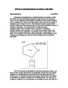
Inside The Mitochondria
Mitochondrion is where the respiration takes place, this is when the glycol sis splits into two carbons and then becomes three lots of carbons this is a series of chemical reaction.
Reference:
http://www.ucl.ac.uk/~sjjgsca/CeelMitochondria.gif
Class notes hand out 12 / 3 / 2008
Website viewed on 20/ 4 / 2008
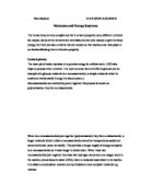
Cardiovascular (heart) System
This picture below is the structure of the heat,
Reference:
Class notes hand out 02 / 04 / 2008
The heart is a muscle that is approximately the size of your fist, it ways round about 1 pound and is located behind and slightly to the left of the breast one this is so the heart can pump blood around the body however the heat pumps about 5 quarters (4.7 litres) of blood everyday or 1800 gallons (6768 litres) of blood everyday.
Reference:
http://www.medtronic.com/heartmc/patient/structure.html
Website viewed on 26 / 4 / 2008
Flow of Blood through the Heart
Reference:
Class notes hand out 02 / 04 / 2008
How the Blood Passes through the Heart
When the blood is deoxygenated it enters through the right atrium and passes through the interior vena cava. The atrium then contrasts and pushes the blood into the right ventricle.
The tricuspid valve is there to make sure that the blood flows the correct way therefore when the right ventricle contracts, it forces the blood through the semi - lunar valve along the pulmonary arteries towards the lungs.
The blood becomes oxygenated within the lungs, the blood then returns to the heat within the pulmonary veins; this then enters the left atrium.
When these contracts it pushes the blood into the left ventricle which has a thick wall because when it contracts it generates high pressure forces the blood through the aorta to the head and the body.
Reference:
Class notes hand out 02 / 04 / 2008
The Cardiac Cycle
Diastole is a period of ventricular relaxation Systole is a period contraction
Normally systole is longer then a diastole
Reference:
Class notes hand out 02 / 04 / 2008
Rate of Heart Beat
In the cardiac cycle the heartbeat on an average is 70 beats per minute (60 seconds) then the time of one beat or one cardiac cycle is 60 divided by 70 seconds is 0.8 seconds therefore you must remember that this is based on the average resting heart rate.
The higher the heart rate is the shorter the cardiac cycle is until a limit is reached when the heart would not have time to fill between successive cycles.
Reference:
Stretch B, BTECH national health and social care, (heinmann 2002).
Cardiac Cycle
The contraction of the myocardium produce pressure changes which results in an orderly movement of the blood, therefore the blood flows from an area of high pressure to an area of low pressure, unless the flow is blocked by the valves.
The event on the right and the left side of the heart are the same, however the pressure on the right side of the heart is lower.
Arial stole is when the heart is full of blood and the ventricle are relaxed therefore both the atria contracts and the blood passes down the ventricle and the ventricular valve opens due to blood pressure.
70% of the blood flows passively down to the ventricles so the atria don't have to contract as much.
Ventricle systole is when the arteries are relaxed and the ventricle wall contracts forcing the blood out, the pressure of the blood forces the atria shut (producing the heart sound 'lub', this sound occurs when the pressure opens the semi - lunar valve and the blood passes into the aorta and the pulmonary arteries.
Diastole is when the ventricles relaxes, however the pressure in the ventricles falls below that in the arteries. When the blood is under high pressure in the arteries courses the semi lunar valve to shut this den produces a second heart sound 'dub', therefore during diastole all the muscle in the heart relaxes, and the blood from the vena cava and the pulmonary vein enters the atria.
The whole cycle starts again.
Reference:
http://www.thestudentroom.co.uk/wiki/Revision_Notes:_The_Heart_Beat
Website viewed on 27 / 4 / 2008
Electrocardiogram (ECG)
Reference:
http://www.highlands.edu/academics/divisions/smpe/biology/th/2122/images/PQRST.jpg
Website viewed on 19/5/08
Electrocardiogram (ECG) is a recording of the electrical changes within the myocardium during a cardiac cycle and consists of a series of three clear waves called deflection waves
P wave is the movement of the depolarization wave from the S-A node through the atria, leading to arterial contraction
QRS wave is ventricular depolarization
T wave is ventricular repolarisation.
Reference:
http://www.highlands.edu/academics/divisions/smpe/biology/th/2122/cvheart.htm
Website viewed on 19/5/08
Blood Vessels
Blood vessels have a simple job and the tube takes the blood around the body therefore the blood goes from the heat, were the blood is pumped to the parts of the body that needs oxygen and takes the blood back to the heart again.
Blood vessels are very valuable because it carries and transport blood, oxygen, nutrients and electrolytes and therefore the vessels also control the temperatures and immunity within the body.
The typical structure is of an inner layer of endothelial cells an example of this is a single layer of cells lining the inside called the tunica intimae.
A muscular layer called the tunica media, and then an outer layer called the tunica adventitia; this contains nerve cells and the blood vessels that supplying the vessel itself!
The tunica adventitia actually contains more connective tissue than anything else, and has fibroblasts in it.
Reference:
(Tucker Louise anatomy and physiology 1988)
http://www.blobs.org/science/article.php?article=54
Website viewed on 28 / 4 / 2008
Arteries
An artery is a blood vessel which leads away from the heart, therefore the pressure within the artery will be very high, however the tunica media of the vessel which hold the muscle is thick because the elastic vessels have to put up with the heart pumping enthusiastically all day long, by stretching to make room for the each swell blood and collapsing once again during diastole.
The artery has a thick wall with a hollow tube; it also has a fibrous outer covering and a middle layer of muscle with an elastic tissue, and the endothelial layer made of the squamous, this is so the artery can carry oxygenated blood from the heart to the body and the pulmonary artery carries deoxygenated blood to the lungs.
Reference:
(Tucker Louise anatomy and physiology 1988)
http://www.blobs.org/science/cells/bvartery.gif
http://resources.schoolscience.co.uk/abpi/heart/heart2.html
Website viewed on 28 / 4 / 08
Capillaries
A capillary are the smallest blood vessel that you find at the end of the bloods rout to the organs this is allowing the gasses (oxygen and carbon dioxide) and nutrients to pass through along with a large amount of water, so that the solution can dissolve in it, which then filters out through the capillary wall and baths the body tissues therefore the liquid is called interstitial fluid.
The ...
This is a preview of the whole essay
Reference:
(Tucker Louise anatomy and physiology 1988)
http://www.blobs.org/science/cells/bvartery.gif
http://resources.schoolscience.co.uk/abpi/heart/heart2.html
Website viewed on 28 / 4 / 08
Capillaries
A capillary are the smallest blood vessel that you find at the end of the bloods rout to the organs this is allowing the gasses (oxygen and carbon dioxide) and nutrients to pass through along with a large amount of water, so that the solution can dissolve in it, which then filters out through the capillary wall and baths the body tissues therefore the liquid is called interstitial fluid.
The capillary job is to distribute (shares out) essential oxygen and nutrients to most parts of the body apart from the deep brain, hyaline, cartilage and the epidermis and the pressure is low.
Reference:
http://www.blobs.org/science/article.php?article=54
Website viewed on 28 / 4 / 2008
Veins
The vain has three layers of the walls through the basic structure to the arteries apart from the veins walls are much thinner and the lumen which is a passage in the centre that carries the blood however this may vary in size, the largest being is the vena cava (from the body into the heart and the pulmonary vain (from the lungs to the heart).
The action of the skeleton muscle pushes the blood through the vessels, therefore the valve within the endothelial layer of the vain to prevent a back flow of blood this is because the blood pressure is very low so the valves are essential (important)
The veins carry deoxygenated blood back to the heart apart from the pulmonary vain.
Reference:
http://www.blobs.org/science/article.php?article=54
Web sight viewed on 29 / 4 / 2008
Blood Pressure
The normal blood pressure is important for a proper blood flow to the body's organ and tissues, therefore each heart beat forces the blood around the rest of the body however the blood moves from high pressure near the heart and low pressure away from the heart.
The force of the blood on the arteries is called blood pressure therefore the blood pressure is determined by the amount of blood pumped by the heart and the diameter of the arteries.
Reference:
Class notes hand out 02 / 04 /08
http://health.howstuffworks.com/high-blood-pressure-in-depth.htm
Website viewed on 29 / 04 08
Measuring the Blood Pressure
Blood pressure is measured as the heart contracts and relaxes
* The systolic pressure is measured when the heart ventricles contracts.
120 mm Hg <-- Systolic
80 mm Hg
* The diastolic pressure is measured when the heart ventricles relaxes.
120 mm Hg
80 mm Hg <-- diastolic
* The normal blood pressure is
20 mm Hg <-- Systolic
80mm Hg <-- Diastolic
In stressful condition can temporarily increase blood pressure.
* High blood pressure is a consistence reading of
140mm Hg
Or higher
90mm Hg
Reference:
http://www.knowledgebase-script.com/demo/article-307.html
http://www.knowledgebase-script.com/demo/admin/attachments/blood-pressure-check.jpg
Website viewed on 29 / 4 / 08
Pulmonary and Systemic Circulation
What is a pulmonary circulation?
* The pulmonary circulation is a circulation of blood from the heart to the lungs and back again.
* The deoxygenated blood travels from the heart to the lungs in the pulmonary artery.
* The blood gets rid of its carbon dioxide (CO2) and replaces it with oxygen (O2), then its returned to the heart passing through the pulmonary vain (from the lungs to the heart) so its ready to be pumped round the body again.
How does this happen?
* Deoxygenated blood from the body enters the right atrium.
* It flows through the tricuspid valve into the right ventricle. The term tricuspid refers to the three flaps of tissue that make up the valve.
* The heart is located roughly in the centre of the chest cavity. It is covered by a protective membrane.
* The pericardium contraction of the ventricle then closes the tricuspid valve and forces open the pulmonary valve.
* Blood flows into the pulmonary artery.
* This branches immediately, carrying blood to the right and left lungs.
* Here the blood gives up carbon dioxide and takes on a fresh supply of oxygen.
* The capillary beds of the lungs are drained by venues that are the tributaries of the pulmonary veins.
* Four pulmonary veins, two draining each lung, carry oxygenated blood to the left atrium of the heart.
Reference:
(Tucker Louise anatomy and physiology 1988)
http://www.bbc.co.uk/science/humanbody/body/factfiles/heart/heart.shtml
Website viewed on 29 / 04 / 2008
What is the systemic circulation?
Systemic circulation supplies (nourishment (food) to all of the tissue located throughout your body, with the exception of the heart and lungs because they have their own systems.
* Systemic circulation is a major part of the overall circulatory system.
* The blood vessels (arteries, veins, and capillaries) are responsible for the delivery of oxygen and nutrients to the tissue.
* Oxygen-rich blood enters the blood vessels through the heart's main artery called the aorta.
* The forceful contraction of the heart's left ventricle forces the blood into the aorta, which then branches into many smaller arteries which run throughout the body.
* The inside layer of an artery is very smooth because it's allowing the blood to flow quickly.
* The outside layer of an artery is very strong because it's allowing the blood to flow forcefully.
* The oxygen-rich blood enters the capillaries where the oxygen and nutrients are released.
* The waste products are collected and the waste-rich blood flows into the veins in order to circulate back to the heart, where pulmonary circulation will allow the exchange of gases in the lungs to take place.
* During systemic circulation the blood passes through the kidneys. This stage of systemic circulation is known as renal circulation.
* During this stage, the kidneys filter much of the waste from the blood.
* Blood also passes through the small intestine during systemic circulation therefore this stage is known as portal circulation, however during this stage, the blood from the small intestine collects in the portal vein which passes through the liver.
* The liver filters sugars from the blood, storing them for later.
Reference:
http://www.fi.edu/learn/heart/systems/systemic.html
http://users.rcn.com/jkimball.ma.ultranet/BiologyPages/C/Circulation.html
Website Viewed on: 16 / 5 / 2008
Structure and the Function of the Blood
What Does the Blood Contain?
The blood it self looks red liquid however if you put it in the test tube it separates the blood in to a pale liquid called plasma and a solid layer of blood.
The blood holds about 55% of plasma and 45% of cells.
Plasma is mostly water with proteins and other chemicals dissolves init, however there are three main type of cells within the blood.
* Red blood cells
* White blood cells
* Platelets
The Structure of the Plasma
* Normal blood plasma is 90-92 % water.
This is the straw-coloured fluid in which the blood cells are suspended, and consists of:
* Dissolved substances including electrolytes such as sodium, chlorine, potassium, manganese, and calcium ions;
* Blood plasma proteins (albumin, globulin, fibrinogen);
* Hormones.
The Function of the Plasma
The medium blood cells are transported around the body (by the blood vessels) and are able to operate effectively; therefore this helps to maintain its optimum body temperature throughout the organism, and helps to control the pH of the blood and the body tissues, maintaining this within a range at which the cells can thrive, this also helps to maintain an ideal balance of electrolytes in the blood and tissues of the body.
Reference:
http://www.ivy-rose.co.uk/Topics/Blood_Structure&Functions.htm
http://www.blobs.org/science/article.php?article=14#2
Website viewed on 16 / 5 / 08
Red Blood Cells Structure
* Biconcave, nucleus - free discs
Function of the Red Blood Cells
* Transport oxygen as ox haemoglobin(for which iron and B12 are required)
* Produces in red blood marrow.
* Give blood its characteristics red colour, thanks to protein called haemoglobin which absorbs the oxygen.
* The lifespan on the red blood cell lasts about 120 days
* The blood cells are then broken down in the spleen and then the liver ( this is where the spare iron is retrieved and recycled)
Reference:
http://www.cancerhelp.org.uk/help/default.asp?page=116#blood
Website viewed on 20/5 /08
The Structure White Blood Cells
* There are two main types of granulocytes and non granulocytes white cells.
* Larger than the red blood cell with a irregular shape and a nucleus
The Function of the White Blood Cell
* To protect the body from infections.
* There are approximately 8000per mm3 in a healthy body.
* Increases rapidly by mitosis in case of serious infection.
Granulocytes
* Defends system against microorganisms an example of this would be viruses and bacteria.
* Granulocytes form 75% of white blood cells
* Attracted by toxins into the tissues and can pass threw the capillary walls.
Non granular leucocytes
* Lymphocytes is when it produces some antibodies, this is formed within the lymphatic tissue and found within all tissues except brain and spinal cord.
* Monocytes eat bacteria and other micro-organisms this is a process which is known as phagocytises.
Reference:
http://www.blobs.org/science/article.php?article=14#2
Website viewed on 19/5/08
Structure of the Platelets
* Small fragile cells with no nucleus
The Function of the Platelets
* Responsible for blood clotting.
* This is formed in red bone marrow.
* 250000 per mm3 of blood.
Reference:
http://www.cancerhelp.org.uk/help/default.asp?page=116#blood
Website viewed on 20/5/08
Respiratory System
Reference
Class handout 16/4/2008
What Is A Respiratory System?
* The respiratory system consist of the nose, mouth, throat, larynx, trachea, bronchi and lungs, therefore the function of the respiratory system is to help gaseous exchange to take place in the lungs and tissue cells of the body.
* Every cell in our body needs oxygen therefore the beating of the heart and the movement of our muscles is by cell division however none of these would have been possible without oxygen.
* One breath is enough for oxygen to entre the body but a deeper breath is more affective, therefore the oxygen within one breath will quickly reach the lungs,
* This will then aboard the vehicles that is specially designed for it.
* The blood cells will spread the oxygen threw out the body and it will represent the source of light for each of the cells within the body because the oxygen is getting to the cells.
Reference:
http://www.ivy-rose.co.uk/Topics/Respiratory_System.htm
http://www.brianmac.co.uk/physiolr.htm
Website viewed on 20/5/08
The Gross Structure of the Repertory System
The nose is an organ on the face which acts as the first passageway for the air to entre the body
The Structure of the Nose
* The nose is made up of cartilage of two nasal bones.
* Its covered with skin, both in side and out side the nose, this is then lined with a mucous membrane which is ciliated for example this has microscopic hairs.
* The two nostrils lead into a bony cavity; this has two chambers, which is divided by a nasal septum.
* The septum is made of the cartilage; therefore the out side of the nose which we can see is mostly up of cartilage as fore the inside of the nose is made up of bone.
* The nasal cavity connects to the Para-nasal sinuses, this is a hollow space inside the bone which is surrounding the nose with full of air and are also lined with the mucus membrane.
The Function of the Nose
The nose is the first organ that the air enters; the nose has three functions,
* To work as an organ of smell
* To moisten and worm the air entering the nostrils
* To filter dust, bacteria and other foreign matter from the air using mucus membrane and its hair.
* The mucus collects any dirt and bacteria and prevents it from passing into the lungs.
* The cilia push the mucus into the throat.
* It is then swallowed and travels to the stomach where any bacteria are neutralised by the gastric acid.
Reference:
http://www.le.ac.uk/pa/teach/va/anatomy/case2/frmst2.html
http://training.seer.cancer.gov/module_anatomy/unit9_4_resp_passages1_nose.html
Website viewed on 20/5/08
Pharynx (Throat)
Once the air has been filtered, moistened and warmed in the nose it travels to the pharynx, a tube which leads from the back of the nose and mouth and divides into the oesophagus (posteriorly) and (anteriorly). It works as part of both the digestive and respiratory system
The Structure of the Pharynx
* The pharynx is about 12.5cm long and is made of muscular and fibrous tissues.
* At the back of the section of the pharynx which connects to the nose are small masses of lymphoid tissue which form the pharyngeal tonsils or adenoids.
* Like the palatine tonsils (at the junctions of the mouth and throat) and pharyngeal tonsils filter bacteria.
The Function of the Pharynx
* It acts as an air passage and also warms and moistens the air.
Reference:
http://www.ivy-rose.co.uk/Topics/Respiratory_Components.htm
http://training.seer.cancer.gov/module_anatomy/unit9_4_resp_passages2_pharynx.html
Website viewed on 20/5/08
Larynx
From the pharynx, air travels down to the larynx (also known as a voice box)
The Structure of the Larynx
* The larynx is a tube positioned between the tongue at the mouth and the trachea (the tube leading to lungs)
* It is made of rings cartilage that is attached to each other by the membrane and ligaments.
* The thyroids cartilage at the top of the larynx, which is a larger in men then in women, this forms the Adam's apple which is often visible in the throat.
The Function of the Larynx
* The larynx is a passageway for air between the pharynx and trachea.
* It filters bacteria, helps in voice production and warms and moistens the air.
Reference:
http://www.ivy-rose.co.uk/Topics/Respiratory_Components.htm
http://training.seer.cancer.gov/module_anatomy/unit9_4_resp_passages3_larynx.html
Website viewed on 20/5/08
Trachea
From the larynx, air travels to the trachea.
The Structure of the Trachea
* The trachea is a continuation of the larynx.
* It is a tube about 10cm long which runs from the front of the neck to the chest were its divided into two bronchi, tubes which lead to the lungs.
* The trachea is made of incomplete ring of hyaline cartilage (anteriorly) and involuntary muscles and connective (posteriorly).
* It is lined with ciliated epithelium which contains mucus-secreting goblet cells.
The Function of the Trachea
* The trachea is a passageway for the air between the larynx and bronchi.
* The goblet secretory cells in the lining secrete mucus which collects any foreign matter or bacteria and the cilia pushes this up towards the larynx.
Reference:
http://training.seer.cancer.gov/module_anatomy/unit9_4_resp_passages3_larynx.html
Website viewed on 21/5/08
Bronchi
The bronchi are the branches of the respiratory tube which transport air in and out of each lung.
The Structure of the Bronchi
* The bronchi connect the trachea to the lungs.
* There are two of them, one on the left and one on the right which enters the lungs at the hilum, a converted depression, where they subdivided into different branches for different lobes of the lungs
* The trachea is made of hyaline cartilage.
* Involuntary muscles and connective tissues and are lined with ciliated epithelium.
The Function of the Bronchi
* To pass air from the trachea into the bronchioles, and to the lungs.
Reference:
http://www.dbcare.org/asthma_files/image001.jpg
http://training.seer.cancer.gov/module_anatomy/unit9_4_resp_passages3_larynx.html
Website viewed on 21/5/08
Bronchioles
The final and finest tube in the passage of the air from the nose and the nose to the lungs is bronchioles.
The Structure of the Bronchioles
* Bronchioles are made of muscular, fibrous and elastic tissues.
* They became progressively smaller as they spread further into the lungs until they are no more than a single layer of flattened epithelial cell (theses look like blood capillary).
* These microscopic tubes are called terminal bronchioles.
The Functions of the Bronchioles
* Bronchioles take the air to the alveoli of the lungs.
Reference:
http://training.seer.cancer.gov/module_anatomy/unit9_4_resp_passages4_bronchi.html
http://www.dbcare.org/asthma_files/image001.jpg
Website viewed on 21/5/08
Lungs
The two lungs are the centre of the respiratory system. It is in these two spongy organs that gases entre and exits the blood.
The Structure of the Lungs
* The lungs are positioned either side of the hart: the left lung is divided into two lobes which are the superior and inferior lobes, as fore the fight lungs is divided into three which are the superior, middle and inferior.
* The lobes are subdivided into lobules.
* The lung tissues are made of bronchioles, alveoli, blood vessels, nerves, connective tissues and elastic tissues.
* They are converted in a special membrane called pleura.
The Function of the Lungs
* The lungs allow the exchange of gases into and out of the blood cells.
Reference:
http://training.seer.cancer.gov/module_anatomy/unit9_4_resp_passages4_bronchi.html
http://www.dbcare.org/asthma_files/image001.jpg
Website viewed on 21/5/08
Pleura
The Structure of the Pleural
* The pleura are a serious membrane that surrounds each lung.
* The pleura has two layers, the inner layer, visceral layer which sticks to the lung tissue and covers the surface and the outer parietal layer which sticks to the chest wall and the top of the diaphragm.
* The two layers are separated by a space called pleural cavity which is filled with a serious fluid.
The Function of the Pleural
The pleural cavity prevents friction between the two layers during the respiration.
Reference:
http://www.cancerhelp.org.uk/cancer_images/Mesolung.gif
http://www.ivy-rose.co.uk/Topics/Respiratory_Components.htm
Website viewed on 21/5/08
How Does the Blood Travel to and From the Lungs?
The pulmonary circulation is the movement of the blood from the heart to the lungs and back.
Ventilation - Inspiration and Expiration
* Air enters the respiratory system.
* This is known as inspiration or inhalation this happens when the pressure is lower inside the lungs and leaves the lungs when the pressure in the atmosphere around the body is lower this is known as expiration or exhalation.
* The action of the muscle is involved in respiration that make theses changes within the pressure and the movement of the air begins.
* The main muscle involved in the mechanics of respiration is the diaphragm which is helped by the intercostals muscle this is positioned between the ribs.
Reference:
Class notes handout
http://www.gcsesciencedoubleaward.co.uk/c3.htm
Website viewed on 24 / 5 / 08
Diaphragm
* The diaphragm is a large muscle.
* It is positioned between the chest and abdomen and separates from each other
The Structure of the Diaphragm
* The diaphragm is made of central sheets of tendon with muscle fibres towards the edges and has three origin which are
> Posterior
> Lateral
> Anterior
* When relaxed it is a dome shape and when its is contracted it is flat
The Function of the Diaphragm
* Inspiration or inhalation happens when the diaphragm contracts, it flattens out and since it forms the bottom of the chesty cavity,
* The cavity then increases within the size and volume, this happens when the pressure is lower inside the chest the air is sucked in because the pressure is lower inside the body than outside.
* Expiration / exhalation happens when the diaphragm relaxes and it becomes a dome shape and pushes up the chest cavity, therefore reducing cavity size and volume which increases the cavity pressure therefore the air rushes out because the pressure is outside the body.
* The diaphragm also helps with expulsive body actions e.g
> Micturition (urine excretion)
> Defecation (faeces expulsion)
> Parturition (giving birth)
Reference:
http://www.gcsesciencedoubleaward.co.uk/c3.htm
http://www.bbc.co.uk/schools/gcsebitesize/biology/humansasorganisms/2breathingrev2.shtml
Website viewed on 24 / 5 08
Alveoli
The exchange of gases in the lungs takes place in the tiny sacs called the alveoli at the end of the terminal bronchioles.
The Structure of the Alveoli
* The alveoli are made of thin layers of squamous epithelial cells and are surrounded by a capillary network.
The Function of the Alveoli
* The exchanges of gases between the circulatory and respiratory systems.
* The pulmonary artery delivers deoxygenated blood to the capillary network which is then oxygenated by contact with air in the alveoli.
* The oxygenated blood then leaves the lungs via the capillary network and the pulmonary vein and travels to the hart to be pumped around the body.
Reference:
http://www.ivy-rose.co.uk/Topics/Respiratory_Components.htm
http://www.dbcare.org/asthma_files/image001.jpg
Website viewed on 21/5/08
What is a Mechanism of Respiration?
* Breathing is a mechanism which enables the entrance and exits of the air into the body as well as the exchange of gases between the blood and the alveoli.
* Gases diffuse from a higher pressure to a lower presser until it's an equal pressure to achieve.
Diffusion
* Diffusion occurs when a strong concentration of a gas comes into contract with a week concentration of the same gas therefore the dissolved gas molecules will move from a strong concentration to a week concentration an till the concentration is week both sides, this is in case of oxygen and carbon dioxide this occurs through the capillary and the alveoli walls.
* The oxygen within the alveoli is under more pressure then the venous, deoxygenated blood in the capillaries so the oxygen passes from the alveoli (high pressure) into the capillaries (low pressure).
* Once the pressure in both is at the same level the exchanges stop.
* The carbon dioxide in the blood is under much more pressure then the carbon dioxide within the alveoli so then it can diffuse through the capillary walls to the alveoli.
* The blood is therefore oxygenated and its waste removed and it now travels back to the heart ready to pump around the body.
* The lungs then expel the carbon dioxide through the process of exhalation.
Reference:
http://arditobook.pbwiki.com/f/626px-Simple_difussion_in_cell_membrane.svg.png
http://www.ebme.co.uk/arts/vent1/index.htm
Website viewed on 22 / 5 / 08
Diffusion of Gases
Once the blood has been oxygenated in the lungs it travels back to the heart and then pumps around the body.
When the blood reaches the various cells of the body, oxygen is then transferred to them by the same method:
The pressure of the oxygen in the blood is high and where as the pressure in the oxygen within the cells is low therefore the oxygen it self passes within the cells.
The amount of oxygen delivered depends on how busy the cell is an example of this would be more oxygen will be delivered to the muscle cell when it is exercising then when it is resting.
The blood delivers oxygen and collects carbon dioxide therefore the pressure within the blood is lower then within the cell so the carbon dioxide passes into the blood and carries it back to the lungs where it will be delivered to the alveoli and then exhaled.
Reference:
http://en.wikipedia.org/wiki/Gas_exchange
http://www.ebme.co.uk/arts/vent1/index.htm
Website viewed on 22 / 5 / 08
Components of Inhale and Exhale Air
* The air that comes into our body contains approximately 21% oxygen and 0.04% carbon dioxide.
* The air that leaves the body contains 16% oxygen and 4% carbon dioxide therefore the air we exhale contains 100 times more carbon dioxide and is also 6% less oxygen then the air we inhale.
Reference:
http://www.concept2.co.uk/training/breathing.php
Website viewed on 25 / 5 / 08
Emphysema and the Surface Area of the Lungs
What is Emphysema?
* Emphysema is a serious lung condition that affects the small air sacs in the lungs (alveoli). This gradual damage to the lungs makes it very hard for you to breath.
* Emphysema is a common condition in smokers and cannot be cured.
What Happens to your Lungs?
* We all breathe in oxygen and breathe out carbon dioxide. This is an essential function of our bodies.
* After air has been inhaled into your lungs, it travels through the bronchial tubes and into your alveoli. The alveoli are small 'balloon-like' structures that are located at the ends of your bronchial tubes.
* It is here, at the alveoli, that oxygen is passed into the blood and carbon dioxide passes back out.
* Emphysema causes the walls of the alveoli to break down so the total surface area available for this gas exchange in your lungs is greatly reduced.
* This means that less oxygen gets into your blood and to your muscles and organs. Also, more of your waste gas (carbon dioxide) stays in your blood because it can't get into the alveoli to be exhaled.
Reference:
http://www.cts.usc.edu/lungvolumereductionsurgery.html
Website viewed on 24 / 5 / 08
The Brains Role in Breathing.
The two centres of the brain are involved in the respiratory centre in the medulla oblongata and the Pons varolii.
* The respiratory centre stimulates inspiration and controls the depth of breathing and its regularity.
* The Pons Varolii stops inspiration therefore provoking expiration. When the respiratory centre tells the diaphragm to contract, air is sucked into the lungs, stimulating nerve cells called stretch receptors found in the tissues.
* The receptors send impulses to the Pons Varolii which then sends impulses to the diaphragm telling it to relax therefore provoking expiration.
Reference:
http://www.nda.ox.ac.uk/wfsa/html/u02/u02_011.htm
Website viewed on 28 / 5 / 08
Digestive System
What is a Digestive system?
The digestive system is a set of organs which transforms whatever we eat into substances that can be used in the body for energy, growth and repair.
Once the food has been broken down by the various chemical processes, and nutrients removed, the rest is excreted as waste.
The whole process involves many different organs and sometimes takes several hours.
What is digestion?
Digestion is a breakdown and transportation of the solid and liquid food into microscopic substances therefore theses substances are then transported into different areas of the body.
There are four stages of digestion:-
* Mouth: ingestion (taking in of food or liquid into the body), chewing and swallowing this is the starch of digestion.
* Stomach: mixing and protein digestion
* Small Intestines: carbohydrates and fat digestion abortion
* Large intestine: waste and excretion
Reference:
http://en.wikipedia.org/wiki/Digestive_system
Website viewed on 28 / 5 / 08
Where Does Digestion Start?
The digestion starts within the mouth where the action of the teeth and saliva combined in the first stage of breakdown which is chewing and partially digesting the food so that it can pass threw more easily along the oesophagus therefore the ball of the food that leaves the mouth is known as bolus.
Saliva
* The salvia is a liquid secreted by three pairs of
* The parotid gland placed below the ear
* The submandibular gland and the sublingual gland both of these are placed below the tongue.
* It contains water, mucus and enzyme salivary amylase.
The Three Functions of the Saliva
* To lubricate the food with mucus, making it easier to swallow
* To start digestion: it contains the enzymes salivary amylase, these acts on cooked starch turning it into shorter polysaccharides.
* To keep the mouth and teeth clean.
Reference:
http://en.wikipedia.org/wiki/Digestive_system
Website viewed 29 / 5 / 08
Enzymes
* Enzymes are an important part of the process, if the digestive system is a conveyor belt then the enzymes are the machine and workers that slowly change whatever is on the belt to make it smaller and smaller so then eventually it can be carried around the body threw the blood.
* The enzymes are made up of protein and acts as a catalysts i.e. they make chemical changes happen in other substances, whilst themselves remaining uncharged.
* They act on food which changes it into small particles.
Reference:
http://scienceaid.co.uk/biology/humans/digestion.html
Website viewed on 29 / 5 08
Tongue
The Structure of the Tongue
The tongue is a muscular organ that is covered with a membrane.
It is held in place by attachments to the mandible (lower jaw) and the hyoid bone.
Tiny projections known as papillae cover the top, increasing its surface area and producing a rough texture.
At the side and the base of the tongue, as well as the palate and at the back of the throat there are special areas known as taste buds.
The Function of the Tongue
* The tongue has tree digestive functions: - taste, chewing and swallowing
* Taste: the tongue is covered with thousands of taste buds which are sensitive to salt, sweet, sour and bitter chemicals within the food and drinks.
* They help us enjoy what we eat and drink and also acts as a first line of defense, warning us when food and drinks are foreign matter are off or unfit for human consumption.
* Chewing: the tongue aid is by moving food around the mouth, pushing it between the teeth and covering it with saliva, which contains enzymes that starts the digestion process.
* The food is turned into a partially digested mass known as a bolus.
* Swallowing: when the food is ready to travel to the stomach, the tongue pushes it to the back of the mouth.
Reference:
http://family.innocentdrinks.co.uk/newsletters/2007/august/
http://www.eatatease.com/themouth.html
Website viewed on 30 / 5 / 08
How does food travel from the mouth to the stomach?
* The action of swallowing and through the tube is known as the oesophagus.
* The tongue pushes the bolus to the back of the mouth, towards the pharynx, muscular tube behind the mouth therefore the food passes into the pharynx and down to the oesophagus.
* The epiglottis is a small flap of cartilage which forms part of the larynx (the windpipe) moves upwards and forwards, blocking the entrance to the larynx.
* This stops the food from going down the wrong way and prevents choking.
Oesophagus
The Structure of the Oesophagus
The oesophagus is a muscular tube which leads from the pharynx, at the back of the mouth, to the stomach, the first main organ of the digestion.
The Function of the Oesophagus
* Carry chewed food from the pharynx to the stomach.
* Food moves along it by muscular concentration this is known as the peristalsis.
* The muscle fibers contract and relax which acts like a wave on the tube which pushes the bolus forwards.
* The lining of the oesophagus secretes mucus to ease and lubricate the passage of the food.
Reference:
http://tabacco.blog-city.com/human_digestive_system_overview__anatomy_function_maintenanc.htm
Website viewed on 30 / 5 / 08
Stomach
The Structure of the Stomach
* The structure of the stomach is J shaped, with an elastic organ which expand and contracts depending on what is in it.
* Food enters from the oesophagus therefore the cardiac sphincter is a valve that stops backflow of the stomach contents and leaves though the pyloric sphincter into the duodenum which is the first part of the intestine.
* The wall of the stomach is a combination of layers of muscle fiber with an inner mucus membrane.
* The latter has lots of folds which is called rugae.
* When the stomach if full they stretch out which enabling expansion then they contract when it empties.
The Function of the Stomach
* Digest proteins threw the actions of enzymes.
* Churns food with gastric juices.
* Helps to lubricate the food by producing mucus (from the mucus membrane)
* Absorbs alcohol
* Kills bacteria by producing hydrochloric acid.
Reference:
http://ezinearticles.com/?The-Stomach-and-Digestion&id=613644
http://www.mydr.com.au/default.asp?article=3351
Website viewed on 31 / 5 / 08
Gastric juices contains
Hydrochloric acid: neutralises bacteria and activates pepsin
Rennin: enzymes that curdle milk protein this only happens in infants.
Pepsin: enzymes that acts on proteins turning them into peptones.
At this stage proteins have been partially digested and, along with the carbohydrates such as starch which were partially digested in the mouth, they have to wait until the small intestine to complete digestion.
Reference:
http://science.jrank.org/pages/2071/Digestive-System-Gastric-juice.html
Website viewed on 31 / 5 / 08
The chemistry of the Digestive System
The digestive process is a combination of different chemical reactions that acts on the food we eat by reducing it to building blocks of nutrients for absorption and to be used by the body.
Every piece of food we eat is composed of fats, carbohydrates (or starch) and proteins.
These must be broken down into relative chemical compounds in order for the body to use them i.e. by time the bread you eat reaches your muscles as energy it has been chewed, churned, liquefied and the starch changed to usable glucose.
What Are Proteins?
* Proteins are made up of one more interlinked polypeptide chains are the building material for the body.
* Protein foods include dairy products, meat, fish and beans. In order to be used by the body they must be converted into a chemical compound called amino acid.
* There are approximately 20 amino acids and two types which is essential and non -essential.
* Essential amino acids are those which the body make itself and should be supplied by dieting
* Non essential amino acids are those which the body can make enough off and are not a dietary requirement.
* Proteins are 1st broken down by pepsin, an enzyme within the stomach, into peptones, which are partially digested protein.
* The peptones are then broken down by the enzymes within the small intestines (trypsin, chmotrypsin) into polypeptides this is a structure formed from the groups from amino acids.
* Lastly, the enzymes peptidase, in the small intestine, breaks the polypeptides into individual amino acid so then it's ready for absorption.
Reference:
http://www.vivo.colostate.edu/hbooks/pathphys/digestion/basics/foodchem.html
http://en.wikipedia.org/wiki/Digestive_system
Website viewed on 1 / 5 / 08
What Are Fats?
* Fats are the body's purest energy source. In order to be absorbed by the body, they need to be converted to fatty acids and glycerol which are compounds that can be absorbed by the small intestines.
* Fats are emulsified by bile salt (which comes from the liver to the duodenum).
* Emulsified fats are broken down into the duodenum by the enzymes lipase which is found within the pancreatic juice and has been converted to fatty acids and glycerol.
* Fatty acids and glycerol are absorbed in the small intestine.
* Emulsify means to turn into emulsion this is a liquid that carries another liquid within it in suspension.
* An emulsified fat is one that has been turned into liquid and carried bile.
* Some paints are known as emulsions because they are exactly that, many medicines include milk of magnesia which is an emulsion.
Reference:
http://www.vivo.colostate.edu/hbooks/pathphys/digestion/basics/foodchem.html
http://en.wikipedia.org/wiki/Digestive_system#Fat_digestion
Website viewed on 1 / 5 / 08
What are carbohydrates?
* Carbohydrate (sugar and starch is found in bread, pasta, biscuits and cakes) these are the energy that is provided for the body.
* They are partially digested in the mouth but are not fully digested until they reach the small intestines.
* Within the mouth the enzymes salivary amylase acts on cooked starch (which are polysaccharides and converts them to shorter polysaccharides this is complex sugar.
* The enzymes amylase converts the polysaccharides to disaccharides in the duodenum.
* The actions of the enzymes maltase, locates and sucrose within the small intestines which then converts and discharges into monosaccharide's which are then absorbed.
Reference:
http://en.wikipedia.org/wiki/Digestive_system#Carbohydrate_digestion
http://www.vivo.colostate.edu/hbooks/pathphys/digestion/basics/foodchem.html
Website viewed on 1 / 5 / 08
Small Intestine
The Structure of the Small Intestine
The small intestine isn't really that small its seven metres long and is divided into three different parts which are the duodenum, the jejunum and the ileum.
The walls have four layers, including a muscular layer, a layer containing blood vessels, lymph vessel, and nerve and an inner mucous membrane.
The inner wall is covered with villi this is a tiny finger like projections which increases the surface area for absorption and contain a network of limp and blood vessels.
The Function of the Small Intestine
The completions of the chemical digestion of food and the subsequent absorption of the nutrients take place within the small intestine.
Nutrients are absorbed threw the villi into the blood and the lymph vessels therefore hardly any food is absorbed elsewhere within the digestive system.
How Does Digestion Absorption Take Place?
* Peristaltic movement mix food with intestinal and pancreatic juices as well as bile.
> The movement push the food against the villi and the intestinal juices are composed of enzymes.
> Maltase, sucrose and lactase which split disaccharides into monosaccharides
> Enterokinase which actives trypsin in pancreatic juice
> Peptidases which split polypeptides into amino acids for example trypsin.
Reference:
http://www.bbc.co.uk/science/humanbody/body/factfiles/smallintestine/small_intestine.shtml
Website viewed on 1 / 5 / 08
*
Absorption: digests food is absorbed threw the walls of the villi:
> Fats, fatty acids and glycerol are passed into the lacteals (lymphatic capillaries).
> These are called lacteals because the fat passes into them in suspension, making the lymph look milky.
> Amino acids and sugar pass along the hepatic - portal vein to the liver.
* The three hormones are in the intestine which help digestion:
> Secretion, pancreozymin and enterocrinin. This stimulates the production of the pancreatic juices, which in turn stimulate the production of intestinal juices.
What Are The Other Functions of The Small Intestines?
* To protect the digestive system from infections
* It is the only section of the digestive system with a direct link to the protective lymphatic system.
Reference:
http://www.bbc.co.uk/science/humanbody/body/factfiles/smallintestine/small_intestine.shtml
Website viewed on 3 / 6 / 08
Large intestine
Structure of the Large Intestine
* The large intestine deals with the waste
* It is about 1.5m long and sits on the draped around the small intestine, in an arch shape.
* It consists of the ceacum, appendix, colon, rectum, anal canal, and anus.
The Function of the Large Intestine
* To reabsorb water and nutrients from the digestive waste to get rid of waste.
* Whenever remains of the food, once it has been thought process of mixing, conversion and abortion carried out in the stomach and small intestines, this is passed into the large intestine.
* Any remaining nutrients are removed and results within the faeces.
Faeces
* Are the unwanted leftovers from the food which then combines with the cellulose (roughage) which is then indigestible this is found in foods like vegetables and bran, dead blood cells bacteria (both living and dead), fatty acid mucus, used to help move the faeces through the large intestines.
* The colour comes from the dead blood cells and bilirubin which is a bile pigment.
Reference:
http://www.malton.n-yorks.sch.uk/ScienceWeb/BiologyWeb/human/digestive_system/kidshealth.html
http://tabacco.blog-city.com/human_digestive_system_overview__anatomy_function_maintenanc.htm
Website viewed on 4 / 6 / 08
Liver
The structure of the liver
* The liver is the largest glands within the body.
* The liver sits at the top of the abdomen, and jus below the diaphragm and just above and to the right of the medial line.
* It is vital because it performs many essential functions.
The function of the liver
* The liver is a vital for cleansing and storage as well as production.
* It removes
* Toxin from drugs, alcohol, and harmful substances.
* Nitrogen from amino acids.
The liver stores
* Vitamin A, B12, D, E, K
* Glycogen is a compound that stores energy
* Iron, from the breakdown of red blood cells and food
* Fats
The liver produces
* Heat this means that the liver is the bodies radiator by producing more heat then any other organs as a result of its various functions.
* Vitamin A (from carotene, found in green leafed vegetables and carrots)
* Vitamin D
* Heparin
* Plasma proteins: albumin, globulin, prothrombin, fibrinogen
* Bile
* Uric acid and urea, from breakdown of red blood cells and de - aminatation of amino acid.
Sonal Mistry