The Pancreas is a large gland that forms part of the Endocrine System, but the Pancreas also has an exocrine part to it.
DISEASES AND DIAGNOSIS Task 1: The Pancreas is a large gland that forms part of the Endocrine System, but the Pancreas also has an exocrine part to it. Despite the fact that it is part of the endocrine system, most of the Pancreas is made up of it's exocrine part. It is this part of the Pancreas that releases a highly Alkali solution that is rich in enzymes into the duodenum via the Pancreatic Ducts. The reason the secretion is of a high pH is because of a high content of bicarbonate Ions and it is these which neutralises the highly acidic clyme as it is moved from the small intestine into the duodenum. The process of the breakdown of Carbohydrates, Lipids, Nucleic Acids and Proteins by Pancreatic Enzymes is called Luminal Digestion. The enzymes trypsin and chymotrypsin are both secreted in inactive form. The enzyme that causes protyrypsin to produce trypsin is called Enterokinase. The Trypsin then goes on to activate prochymotrypsin to form chymotrpysin. This process is carried out to prevent the Pancreas from digesting itself. The other pancreatic enzymes are secreted in active form. The secretion of Pancreatic enzymes is a continuous process. To promote the secretion of the enzymes, endocrine cells in the duodenum release the hormone Secretin and this hormone also promotes the secretion of a fluid, which is copious, watery and rich in bicarbonate. (Information from

The structure and function of Proteins
The structure and function of Proteins This essay clearly divides into two sections: structure, and function. Hence I will begin by talking about the structure of Proteins and will go on to talk about their functions. The building blocks of proteins are "amino acids", which differ to other molecules due to their nitrogen content. The basic amino acid looks like this, with a line of N-C-C. The N has two H atoms bonded to it, while the end C atom has a double bond to an oxygen atom and has a hydroxyl group bonded to it. The middle carbon atom bonds with a H atom, and has a "variable side group" attached onto it. Amino acids are very soluble, principally for the reason that the Oxygen and Nitrogen in them is very electronegative, meaning that the amino acids are readily carried around in aqueous state. Amino acids are "amphoteric" meaning that in different circumstances they can act as both acids and alkalis. When in a specifically acidic or alkaline solution, the amino acid forms a "zwitterion": When added to an acid, the zwitterion form of the amino acid acts as a buffer by taking up H+ ions to keep the concentration oh hydrogen constant, and hence to raise the pH. However, when in an alkaline solution, the amino acid acts as a buffer in a different way. It gives away a H+ ion to the solution, to lower the pH and hence it controls the concentration of Hydrogen ions. The
Describe the structure of the liver and explain its
Describe the structure of the liver and explain its functions in maintaining homeostasis The liver is the largest organ in our body it weighs about 1.5kg, it is situated in the abdomen just below the diaphragm. The liver helps to regulate the composition of the blood, it has two blood supplies the hepatic artery and the hepatic portal vein and also has a blood removal hepatic vein. The hepatic artery carries oxygenated blood from the dorsal aorta, whilst the hepatic portal vein carries deoxygenated blood from the gut. The hepatic vein is responsible for carrying deoxygenated blood from the liver to the inferior vena cava; these blood vessels are known as the interlobular vessels. The structure of the liver consists of a large number of lobules, these are cylindrical in shape and each one is approximately 1mm in diameter. Each lobule consists of liver cells arranged in rows radiating from the centre towards the margin. Liver cells are similar although they have many different functions; however they have large numbers of mitochondria and a large Golgi Apparatus. In the centre of each lobule there is a branch of the hepatic vein this is known as the central or intralobular vein. The interlobular vessels are connected to the central intralobular vessel using a system of capillary like sinusoids, they run parallel to and also come into contact with, the chains of liver

Structure and function of the digestive system.
STRUCTURE AND FUNCTION OF THE DIGESTIVE SYSTEM The function of the digestive system is to break down large molecules in food, such as protein and starch, into monomers that can easily be absorbed into the body and with a balanced diet it does its job efficiently. The alimentary canal is a long coiled tube that runs through the body from the mouth to the anus. It consists of associated glands, the salivary glands, the liver and the pancreas. This muscular tube is organised into several distinct regions. The overall process of nutrition can be divided into several stages. Ingestion is taking in food. Humans put food into the mouth where it is chewed. Swallowing takes it down through the oesophagus and into the stomach. Peristalsis, rhythmic contractions of the gut wall, propels the food through the alimentary canal. Mechanical breakdown is how food is broken down into smaller pieces by chewing in the mouth and the churning action of the stomach. Digestion is the complex breakdown of foods such as carbohydrates, lipids and proteins into simpler molecules. Absorption is where simple food molecules pass into he blood stream in forms such as amino acids, fatty acids, vitamins, minerals, sugars and water. Egestion is where undigested food is eliminated from the body via the act of defecation. A balanced diet is required to give the human body all the nutrients it needs
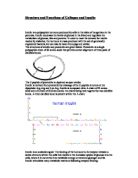
Structure and Functions of Collagen and Insulin.
Structure and Functions of Collagen and Insulin Insulin is a polypeptide hormone produced by cells in the islets of Langerhans in the pancreas. Insulin decreases the levels of glucose in the blood and regulates the metabolism of glucose, fats and proteins. In order to meet the demand for insulin needed by diabetics, the hormone is mass-produced with the aid of genetically engineered bacteria, but can also be taken from pigs and cattles. The structures of insulin and proinsulin are given below. Proinsulin is a single polypeptide chain of 86 amino acids that permits correct alignment of three pairs of disulfide bonds. The C-peptide of proinsulin is depicted as open circles. Insulin is derived from proinsulin by cleavage of the C-peptide structure at the dipeptides Arg-Arg and Lys-Arg. Insulin is composed of an A chain of 21 amino acids and a B chain of 30 amino acids, the chains being held together by two disulfide bonds. A third disulfide bond is present within the A chain. Insulin is an anabolic signal. The binding of the hormone to its receptor initiates a series of events within the cells that results in the increased uptake of glucose into the cells, where it is converted into metabolic energy or stored as glycogen and fat. Insulin stimulates many metabolic reactions following receptor binding: * stimulates glucose transport * stimulates amino acid transport

Describe the gross structure and functions of the main body systems. P3
Describe the gross structure and functions of the main body systems. P3 Introduction I will try describing the gross structure and also the functions of the 10 systems in our body. A system is made up of different tissues and organs working together to perform a specific function in the body. Cardiovascular System The cardiovascular system comprises the heart and blood vessels that circulate blood throughout the body, bringing oxygen and nutrients to muscles and organs and then returning it to the heart to be pumped again. The cardiovascular system includes: * The heart: the pumping organ * Blood vessels : a closed circuit of tubes * Arteries : carry blood to all parts of the body * Capillaries : carry blood from arteries to veins * Veins : carry blood back to the heart * Blood : consisting of liquid plasma and various blood cells Position of the heart The heart is a muscle about the size of a person's fist in the middle of the chest, tilted toward the left side, just under the sternum in a place called the mediastinum. Respiratory system The function of the respiratory system is to transport air into the lungs and to facilitate the diffusion of Oxygen into the blood stream. Its also receives waste Carbon Dioxide from the blood and exhales it. The respiratory system comprises of the nose, mouth, throat, larynx, trachea, bronchi and lungs. Oxygen is
Management style, culture & organizational structure.
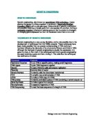
GENETIC ENGEERING GENETIC ENGEERING Genetic engineering, also known as recombinant DNA technology, means altering the genes in a living organism to produce a Genetically Modified Organism (GMO) with a new genotype. Various kinds of genetic modification are possible: inserting a foreign gene from one species into another, forming a transgenic organism; altering an existing gene so that its product is changed; or changing gene expression so that it is translated more often or not at all. TECHNIGUES OF GENETIC ENGEERING Genetic engineering is a very young discipline, and is only possible due to the development of techniques from the 1960s onwards. These techniques have been made possible from our greater understanding of DNA and how it functions following the discovery of its structure by Watson and Crick in 1953. Although the final goal of genetic engineering is usually the expression of a gene in a host, in fact most of the techniques and time in genetic engineering are spent isolating a gene and then cloning it. This table lists the techniques that we'll look at in detail. TECHNIQUE PURPOSE Restriction Enzymes To cut DNA at specific points, making small fragments DNA Ligase To join DNA fragments together Vectors To carry DNA into cells and ensure replication Plasmids Common kind of vector Genetic Markers To identify cells that have been transformed PCR To

Unit 5 P3: Outline the gross structure of all main body systems
Jessica Bascombe P3: Outline the gross structure of all main body systems Digestive system: This is made up of the gastrointestinal tract also called the digestive system and the liver, pancreas and gall bladder. The gastrointestinal tract is a series of hollow organ that joined in a long twisting tube from mouth to the anus; the hollow organs made up the GI tract are the mouth, oesophagus, stomach, small intestine, large intestine which includes the rectum and anus. Food enters the mouth and passes to the anus through the hollow organs of the GI tract the liver, pancreas and goal bladder are the solid organs of the digestive system. The digestive system helps the body digest food, this is important for breaking down food into nutrients which the body uses for energy growth and cell repair food and drink must be changed into smaller molecules from nutrients before the bloody absorbs them and carries them to the cells throughout the body, the body breaks down the nutrients from food and drink into carbohydrates, protein, fats and vitamins. Digestion works by moving food through the GI tract, digestion begins in the mouth with chewing and anus in the small intestine as food passes thought the GI tract it mixes with digestion juices, which causes the large molecules of food to break down into smaller molecules the body then absorbs these smaller molecules through the walls of
current structure and original structure
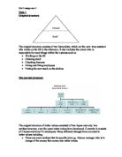
Task 1 Original structure The original structure consists of two hierarchies, which are the part- time assistant who works on the till in the afternoon. It also includes the owner who is responsible for most things within the business such as * Working on the till * Ordering stock * Checking finances * Hiring and firing employees * Putting the new stock on the shelves. The current structure The original structure of better values consisted of two layers and only two workers however, over the years better values have developed. Currently it consists of 3 layers and about 23 employees. Many different changes have occurred to better values including * There are more workers that do specific jobs e.g. finance manager who is in charge of the money that comes into better values. * There are more layers in the current hierarchical structure, which means that jobs have become more specialised. * In the original structure there, where only two workers this means that it was easier for the owner to communicate with his employee. Whereas in the current structure there are more employees and three different levels. This means that it could be harder for the employer to communicate to his staff. In the current structure, all the employees have specific job roles the bottom layer of employees is self-explanatory. The middle layer consists of the store manager. They are in

Structure of the Alimentary Canal in relation to digestion and absorption
Structure of the Alimentary Canal in relation to digestion and absorption There are two stages in human digestion: . Mechanical breakdown-the large particles of ingested food are broken down into smaller pieces by the teeth in the mouth. 2. Chemical breakdown - the large molecules of food are hydrolysed (broken down) by digestive enzymes into smaller, soluble molecules. The alimentary canal (human gut) has the same general structure along its whole length but in some areas, it is specialised to carry out various roles. It extends as a tube from the mouth to the anus and along its length, the wall is composed of four layers: . Mucosa- This is the innermost lining of the gut wall. It lubricates the passage of food with mucus and also protects it from the digestive action of enzymes. The mucosa surrounds the lumen which is made up of glandular epithelium and connective tissue containing blood vessels and lymph vessels. 2. Submucosa- This is a layer of connective tissue that contains nerves, blood vessels and lymph vessels together with elastic fibres and collagen. 3. Muscularis Externa- This is made up of circular and longitudinal layers of smooth muscle fibres which control the shape and movement of the gut. 4. Serosa- This is the outermost layer and is made up of loose connective tissue which provides protection from friction against other organs. Different parts


