IT Key Skills In Biology
Introduction:
The purpose of this experiment is to plan design and carry out an experiment to compare the stomatal density of the upper and lower side of a leaf of my choice.
This is relevant to my AS course in Biology as I am currently studying transpiration. This is a process involoving stomata opening and closing to release moisture from the plant. Therefore the amount and distribution of these stomata is very relevant to what I am learning.
Null Hypothesis is that ‘there will be no difference between the stomatal density on the upper and lower sides of the leaf.
Apparatus:
- leaves of a privit or similar plant
- plain glass slides and cover slides
- scalpel/sharp knife
- nail varnish
- Electron Microscope
- Small painting brush
- Forceps
Method:
Method for determining stomatal density:
(see attached sheet for source of method)
- Select a few leaves from a privit or similar plant
- Cover the upper surface of one of the leaves with a thin layer of nail varnish using a small brush
- Repeat this on the lower surface of one of the other leaves
- When varnish is dry peel layer off upper surface of the first leaf. (Use forceps)
- Place this on a microscope slide with a tiny amount of water and label the slide U (upper surface)
- Repeat this process with the lower surface of second leaf. (label this slide L)
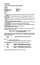
- Examine both slides under microscope using high and medium power.
- Take an area on each leaf to count stomata. Calculate the area of the field of view. ( formula = πr²)
- From this calaculate the number of stomata per unit area on each side of the leaf.