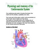
Some of the more important anatomical structures of the heart:
Right atrium: Receives deoxygenated blood into its upper posterior corner from superior vena cava, Inferior vena cava and coronary sinus open into its lower posterior corner.
From the right atrium blood empties into the right ventricle through the atrioventricle valve, Blood flow is in horizontal & forward direction from right atrium to right ventricle, because right ventricle is situated in front as well as to the left of the right atrioventricular opening.
Tricuspid valve: Blood flow from the right atrium to right ventricle through the right atrioventricular orifice which is guarded by the tricuspid valve.
Right ventricle: Walls of right ventricle are thicker then those of the right atrium and possess muscular ridges called trabeculae carneae.
Pulmonary valve: consist of three semilunar cusps, this valve is closed during ventricular relaxation (diastole), preventing backflow of blood from the pulmonary trunk into the right ventricle.
Left atrium: receives oxygenated blood from the lungs, blood moves from the left atrium to the left ventricle from where it is then pushed out to supply oxygen rich blood to rest of the body. Left atrium is divided into two portions: A posterior half and an anterior half
-Posterior half has smooth walls and is the inflow part of the chamber, four pulmonary veins empties into this half
-Anterior half walls have musculi peatinat on them
Mitral valve: Also known as bicuspid valve because it possesses two cusps. it prevents reflux during ventricular contraction.
Left ventricle: Lies to the front of the left atrium, blood flow into this chamber of the heart is in a forward direction. Myocardium is the thickest in the walls of this chamber, up to three times thicker then the walls of the right ventricle.
Aortic valve: Consist of three semilunar cusps, prevent backflow of blood from ascending aorta during ventricular diastole
Right pulmonary artery: Slightly longer and larger then the left coronary artery. It emerge from behind the superior vena cava and runs horizontally to the right.
Left pulmonary artery: Shorter and smaller then the right pulmonary artery, it emerge from within the concavity of the aortic arch and runs horizontally to the left.
Pulmonary veins: Returns oxygenated blood to the left atrium, usually there are four pulmonary veins two from each lung
Aorta: Is the biggest artery in the body that takes oxygenated blood directly from the left ventricle. It divides into ascending and descending aorta and then further divides into different branches
Superior vena cava: Formed by the union of the two brachiocephalic veins, it terminates into the right atrium
Inferior vena cava: Arise from the level of 5th lumber vertebral vertebra, and bring blood back to right atrium from the lower body.