Why and When Is UltraSound used In Pregnancy?
Ultrasound is considered to be safe and an accurate investigation into the fetus. Diagnosis can be spotted early and confirmation of early pregnancy. Ultrasound can observe and measure the embryo and find the exact date of pregnancy. A visible heartbeat can be seen and detectable by pulsed Doppler ultrasound by about 6weeks. Missed abortions and will usually give pictures of an absence of fetal poles or heartbeat. Fetal body measurements are made to identify the gestational age of the fetus.
The Following Measurements Are Usually Made:
The crown-rump length- which can be made between7-13 weeks and can give an accurate estimation of the gestational age.
The Biparietal Diameter- is between two sides of the head and is usually measured in 13weeks. From 13 weeks it normally starts at 2.4cm and increases to about 9.5cm
The Femur Length- measures the longest bone in the body and this reflects on the longitudinal growth of the fetus.
The Abdominal Circumference- This is a measurement made during late pregnancy. It has more of an effect on fetus size and weight.
Summary of Ultrasound
Ultrasound is now something for today and for the future. It was first brought in 1950 and is now used in almost all Hospitals it can be used on pregnant women, which is an advantage to their health and to their child’s because diagnosis can easily be detected at an early date. Ultrasound can also be used for particular illnesses like the treatment of kidneys the shock wave can shatter the stones in your kidney into tiny pieces. The limitation of ultrasound though is safety because there are ill effects such as low birth weight, speech and hearing problems. The argument though is that ultrasound is now a more 21st century technology and is becoming safer gradually. It is more accurate when it comes to assessing and monitoring pregnant women. For the foreseeable future it looks like ultrasound will continue to be used and improvements to better it will always be made.
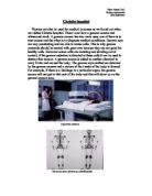
As a part of my course work, I visited the Physics Department at Queen Elizabeth Hospital in Selly Oak, to identify the usage of physics in hospitals and how it benefits the human body.
During my visit I was shown different machines using different aspects of physics.
Ultrasound scanning, This works by taking a 2d image of the foetus or organ, these machines take several images then convert them to a 3d image using specialised computer software. With a continuous picture of the moving foetus can be depicted on a monitor screen. Very high frequency of sound waves of between 3.5 to 7.0 megahertz are used.
They are emitted from a transducer which is placed in contact with the maternal abdomen and is moved to look at any particular content of the uterus. Repetitive arrays of ultrasound beans scan the foetus inn thin slices and are reflected on the same transducer.
How is this benefiting?
The information obtained from different reflections are recomposed back into a picture on the monitor. Movements such as foetal heart beats or malformations of the foetus can be seen. All this can be assessed and measurements are made accurately on the images displaced on the screen. Such as measurements of size and growth in the foetus.
Why and when is ultra sound used in pregnancy.
Ultra sound scan is currently considered to be safe it is accurate and very cost effective investigation of the foetus. This plays an important role in pregnant women.
What about safety?.
It has been over 40 years since ultra sound was first used on pregnant women. Large amount of studies show that apparent ill effects such as low birth weights speech and hearing problems brain damage and non right handedness is reported. The complexity of some of the studies have made the observations difficult to interpritate. Every now and then ill effects of ultrasound on the foetus appears as news items. The main areas of concern are the use of pulsed Doppler.
Doppler Ultra sound.
Doppler ultrasound is based upon the Doppler Effect. When the object moves ultra sound waves are moving it changes the frequency of the echoes. Doppler ultrasound measures the change in frequency of the echo and how fast the object is moving. This is only used for the rate of blood flow through the heart and major arteries.
Frequency is measured in Hertz, the abbreviation for Hertz is Hz a frequency of 1 Hz is one vibration per second if a ruler vibrates a hundred times in one second the frequency is 100 Hz.
Frequency in Hz = Number of oscillation or vibrations / Time taken in seconds.
Kidney stones are hard deposits which can build up inside a person’s kidneys. They can be painful and dangerous and are sometimes removed by surgery. Another treatment is to direct ultrasonic (very fast) vibrations at them. This breaks the stones into tiny pieces which then pass out of the kidney in the urine.
Ultrasound can be used to break down stone and weld plastic. At low intensities the vibrations can be used to shake dirt out of complex shapes such as jewellery.
Radiation Therapy
Shortly after the discovery by the German Physicist Wilheim Roentgen in 1895, the powerful rays are being used to effectively treat cancer. Today an increasing number of patients have their cancers treated successfully with a few side effects and preservation of normal tissue using radiation therapy.
What is Radiation Therapy?
About 50-60% of cancer patients are treated with radiation at some time during their disease. Radiation therapy is the careful use of high radiation to cure cancer or to relieve a cancer patients pain. Radiation Therapy works because the radiation destroys the cancer cells ability to re-produce and the body naturally gets rid of these cells. Nuclear medicine is the subspecialty within the field of radiology comprising diagnostic examinations that result in images of the body anatomy and functions. The images are developed based on the detection of the energy emitted from a radio-active substance given to the patient either intravenously or by mouth. Generally radiation to the patients is similar to the resulting from standard x-ray examinations.
Some of the Common uses of the procedure.
Analysis kidney function
Image blood flow and the function of the heart
Scan lungs for respiratory and blood flow problems
Locate the presence of infection
What Does the Equipment Look like
As with most nuclear medicine examinations, patients lie on a scanning table. The patient can only see the specialised nuclear imaging camera used during the procedure which is enclosed in metallic housing designed to facilitate specific parts of the body’s images. It can look like a large round metallic apparatus suspended from a tall moveable post or a metal arm which hangs over the examination table. The camera can also be similar to a computed tomography (CT) scanner which looks like a doughnut or the camera is beneath the table. The data is processed from a computer nearby or in another room.
How does the Procedure Work
A small dose of radioactive material called a radiopharmaceutical or tracer, is intravenously or orally localised on the infected body organs. The compounds job is to collect in the organ and release energy as gamma rays The gamma camera detects the rays and produces the images with measurements or organs and tissues onto a computer.
How the Procedure is Performed
A radiopharmaceutical is usually given intravenously, into a vein. Depending on the type of scan the imaging can be immediate, a few hours later or even several days after the injection. The imaging times vary from 20 to 45 minutes. The radiopharmaceutical used is determined by the area under investigation because some compounds work better in specific organs than others. Depending on the scan it can take seconds or several days for the compounds to travel through the body and reach it’s target. A physician specialised in this area will check the quality of images to ensure an optical study has been performed, the images are clear.
Patients experience during the Procedure
Some minor discomfort may arise during the intravenous injection from the needle or in cases of special studies when a catheter is placed into the bladder. Most of the radioactivity, compound, passes out of the body through urine or stools with the rest lost over time through natural loss.
Benefits versus Risks
Benefits
The information provided by nuclear medicine examinations is unique and not available by other imaging procedures. For many diseases this is the only quick and reliable sources of examination to provide the necessary treatment. This is also less traumatic for the patient than exploratory surgery and allergic reactions are extremely rare.
Risks
Because the doses of radiopharmaceutical given are very small, nuclear medicine procedures result in exposure to small doses of radiation. Nuclear medicine has been used for more than five decades and there are no known long-term adverse effects from such low-dose studies. Radiopharmaceutical treatment is harmful to the foetus, therefore, during pregnancy the mother is given this treatment minimally such as in an ultra-scan. Limited doses and usage on children and finally as in all cases the risk of allergic reactions to anything administered to the body which it cannot produce naturally.
Radiation is something which is sent out or radiated from an object. Towards the end of the 19th Century a French scientist, Becquerle, discovered that uranium gave out radiation. The radiation blackened a photographic plate. Radiation can be detected because it causes ionisation. Humans have no sense which can detect ionising radiation. We must use instruments to detect it. Ionising radiation ionises atoms as the radiation passes through material it removes electron from atoms producing ions. Ions unlike atoms have a charge, by detecting this charge we can detect ionising radiation.
Alpha, Beta and Gamma Radiation
There are three different types of ionising radiation which can be emitted by radioactive isotopes.
The environment around us is permeated by background radiation which is nuclear radiation from various sources. Cosmic radiation mainly from the sun has always been present as radiation from rocks such as granite. More recently background, radiation has come from testing of atomic bombs and catastrophic accidents in nuclear power stations.
Basics of Radiation
Characteristics of Alpha Radiation
- Alpha radiation is not able to penetrate the skin
- Alpha-emitting materials can be harmful to humans if the materials are inhaled, swallowed or absorbed through open wounds
- A variety of instruments have been designed to measure alpha radiation. Special training in using these instruments is essential for making accurate measurements
- A civil defence instrument (CD V-700) cannot detect the presence of radioactive materials that produce alpha radiation unless the radioactive materials also produce beta and/or gamma radiation
- Instruments cannot detect alpha radiation through even a thin layer of water, blood, dust, paper or other materials because alpha radiation is not penetrating
- Alpha radiation travels a very short distance through air
- Alpha radiation is not able to penetrate turnout gear, clothing or a cover on a probe. Turnout gear and dry clothing can keep alpha emitters off of the skin.
Characteristics of Beta Radiation
- Beta radiation may travel meters in air and is moderately penetrating
- Beta radiation can penetrate human skin to the ‘germinal layer’ where new skin cells are produced. If beta-emitting contaminants are allowed to remain on the skin for prolonged period of time, they may cause skin injury
- Beta-emitting contaminants may be harmful if deposited internally
- Most beta emitters can be detected with a survey instrument (CD V-700 provided the metal probe cover is open). Some beta emitters produce very low energy, poorly penetrating radiation that may be difficult or impossible to detect such as carbon-14, tritium and sulfur-35
- Beta radiation cannot be detected with an ionisation chamber such as a CD V-715
- Clothing and turnout gear provide some protection against most beta radiation. Turnout gear and dry clothing can keep beta emitters off of the skin.
Characteristics of Gamma Radiation
- Is a form of electromagnetic radiation and not a stream of particles,
- It is not deflected by electric or magnetic fields because it does not carry a charge
- Has very high energy
- It can travel a very long way in the air even passing through several centimetres of lead or an even thicker piece of concrete
- The following is a formulae for gamma radiation
The energy of the emitted gamma photon is about 140000 eV (1 eV = 1.6 x 10 –19 J)
Development for the future
3-D Ultrasound is quickly moving our of the research and development stages and is now employed in a clinical setting. It is faster and more advanced with commercial module coming into the market. The scan requires special probes and software to accumulate, gather, and render, produce, the images and the results time is vastly reduced from minutes to fractions of seconds.
A good 3-D image is often very impressive to the parents And further 2-D scans may be extracted from 3-D blocks of scanned information. Volumetric measurements are more accurate and both doctors and parents can better appreciate a certain abnormality or the absence of a certain abnormality in a 3-D scan than a 2-D scan one and there is the possibility of increasing psychological bonding between the parents and the baby.
An increasing volume of literature is building on the usefulness of 3-D scans and the diagnosis of congenital anomalies could receive revived attention. bg Evidence has already suggested that smaller defects such as spina bifida, cleft lips/palate and polydactyl may be more lucidly demonstrated. Other more subtle features such as low set ears, facial dysmorphia or clubbing of feet can be better assessed leading to more effective diagnosis of chromosomal abnormalities. The ability to produce good pictures is still dependant on operator skills, the symniotic fluid around the foetus it’s position and the degree of maternal obesity so that a good image is not always readily obtainable. Most recently 4-D or dynamic 3-D scanners are on the market resulting in reports of it’s importance in mother and baby bonding and the attraction of seeing the baby’s face and movements before birth.
Researching the area nuclear physics has broadened my understanding of the word ‘nuclear’. As a I child I have only been told it’s uses as a bad thing such as nuclear weapons, a powerful gas which kills all living organisms. The use of nuclear in medical developments is not only developing in safer use but also for advanced cure of many developing diseases, to try and detect them in early stages. ‘The Enemy is your Friend’.