Tahir Ahmed L6G
Physics coursework
Mrs Boardman
Christie hospital
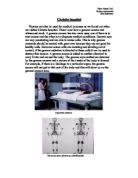
Physics can also be used for medical purposes as we found out when we visited Christie hospital. There I saw how a gamma camera and ultrasound work. A gamma camera has two main uses; one of them is to treat cancer and the other is to diagnose medical conditions. Gamma rays are very penetrating and are able to ionise cells. This is why gamma materials should be treated with great care because they are not good for healthy cells. However cancer cells are mutating and dividing out of control, if the gamma radiation is directed at these cells it can be used to destroy this tumour. A gamma source is added to another chemical to carry it into and around the body. The gamma rays emitted are detected by the gamma camera and a picture of the inside of the body is formed. For example, if there is a blockage in a particular organ, the gamma source will not get to that part of the body and this will show up on the gamma camera scan.
A gamma camera
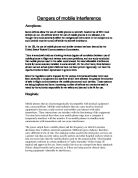
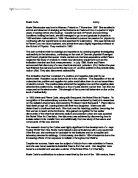
The bone scan shows up arthritic joints
Patients who are going to have this sort of treatment are injected with a radioactive drug depending on
- The type of radiation
- The energy of the radiation
- The organ to be studied or treated
- The rate of radioactive decay
- The rate of clearance from the body
The most commonly used isotope is technetium-99m, which emits gamma rays with energy of 140keV. This isotope has a half-life of 6 hours, which is useful for scanning because the radioactive material will not stay in the body for long. If it did stay in the body for long then this would cause more harm to the patient than help. Cells with cancer absorb more of the radioactive isotope than normal cells can since they use up the isotope at a much more rapid rate (cancerous cells use up glucose approximately 20 times that of a normal tissue). Using cyclotran devices produces radionuclides; a cyclotran is a particle accelerator, which bombards the nuclei of stable atoms. The charged particles must have enough kinetic energy to overcome the repulsive effects of a positively charged nucleus. The radionuclides emit faint gamma rays, which are then measured by the gamma camera. The gamma camera has a large crystal detector (called a scintillation crystal). These crystals detect the emitted radiation signal and convert that signal into faint light. The light is then converted to an electric signal, which is then digitised (converted into a computer signal) and reconstructed into an image by a computer. The resulting image is viewed on the system monitor. An important part of the gamma camera is the collimator because it selects the direction of the incident photons.