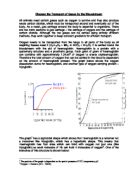
The graph has a sigmoidal shape which shows that Haemoglobin is a tetramer not a monomer like Myoglobin, which has a hyperbolic shape. This means that Haemoglobin has four sites which can bind with oxygen not just one (like Myoglobin) so each molecule of Hb can hold 4 molecules of oxygen. One of the branches of this structure is shown below:
The significance of the graph is that oxygen will be released from the protein at a low pressure only - when PO2 is high, as in the capillaries of the lungs or in the heart, oxygen binds with haemoglobin, but when PO2 is low, as in tissues, oxygen is released from Hb. This is useful as it means that oxygen will be released in areas that need it most such as in muscles not in areas or high blood pressure such as the heart which do not need huge amount of oxygen to be released.
There are other types of oxygen carrying proteins such as foetal haemoglobin and myoglobin. Foetal haemoglobin has a higher affinity for oxygen than normal haemoglobin which is important so that mother’s blood brought to the foetus will be able to be taken by the foetus’ blood when the two types of haemoglobin meet. Myoglobin is found in muscle tissue and takes up oxygen more readily than normal haemoglobin so that the Myoglobin can act as a temporary store for oxygen inside muscles.
In all types of oxygen carrying proteins, the presence of carbon dioxide decreases affinity for oxygen. In terms of the graph, the line would be shifted to the right – this is known as the Bohr Shift. The Bohr shift occurs due to the decreased pH of the blood when carbon dioxide is present. In biological terms, this means that the more carbon dioxide present, the more oxygen is released.
Carbon dioxide levels most be controlled in order to keep a constant blood pH level. CO2 is carried around the blood using three methods. Firstly, approximately 7% of the gas can be dissolved in the plasma section of blood in water. Secondly, 23% can be combined with proteins. Finally, 70% of carbon dioxide is carried in hydrogen carbonate ions in red blood cells – this is the most important method of transport.
The first method is quite simple and simply involves the dissolving of the gas into the plasma, which lowers the pH of blood (as carbon dioxide + water → carbonic acid – a weak acid) thus creating the Bohr shift. This is not the main method for transporting carbon dioxide as it is very slow (although it can be accelerated to 5000x by carbonic anhydrase but this only occurs in red blood cells)
The second method involves combining of carbon dioxide and haemoglobin to form carbamino haemoglobin.
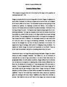
The third method is the most complex but is the bodies preferred method of carbon dioxide transfer. The diagram below represents this transfer method:
In the diagram we can see that the carbon dioxide produced by the cell from respiration (C6H12O6 + 6O2 → 6CO2 + 6H2O) diffuses out of the cell and into a red blood cell in a neighbouring capillary (and about 7% does not reach the cell but instead is dissolved in the blood plasma). Inside the cell, 23% of the carbon dioxide combines with haemoglobin to produce carbamino haemoglobin. The remaining 70% is dissolved in water at a rate 5000 times faster than normal with the aid of the enzyme carbonic anhydrase. The hydrogen carbonate then dissociates into hydrogen carbonate ions:
CO2 + H2O → H+ + H2CO3
The H+ then combines with Haemoglobin to produce H2Gb. This occurs because proteins are made of amino acids which have a buffer effect and combine with H+ ions. The remaining HCO3+ ions diffuse out of the cell and water and Cl- ions also diffuse in to balance out charges.
In conclusion, oxygen is transported around the body using the protein Haemoglobin. There are various types of haemoglobin but they all have similar uses. The structure of these proteins allows them to release oxygen at low partial pressures only, so it is released in areas that need it most. Various factors including carbon dioxide presence can affect haemoglobin’s affinity for oxygen. Carbon dioxide is transported round the blood using 3 main methods – the main method involves storage in red blood cells as hydrogen carbonate ions.
The position of the graph is dependant on the partial pressure of CO2, temperature, pH.
Oxygen = Diatomic (O2 – HbO8)