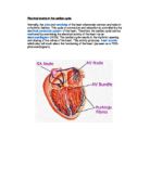
The heartbeat is initiated in a specialised area of muscle in the right atrium called the sinoatrial node (SAN) or the pacemaker. The SAN starts the waves of depolarisation, which results in contraction.
Although other cells in the heart are also capable of emitting impulses spontaneously, the sinus node has the most frequent inherent rate of firing. Consequently, it serves as the dominant pacemaker of the heart.
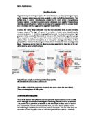
The electrical impulse emitted by the SAN spreads to sarcomeres (cells that have the capacity to shorten or contract. They are the basic muscle fibres of the heart) in the surrounding right atrium. From there, the impulse propagates from one cell to another until the entire myocardial wall of both atria is depolarized. The general direction of the wave front created is from right to left and from the upper atria downward
The waves spread out over the 2 atrial walls so that they contract. There is a band of fibres between the atria and ventricles, which have a high electrical resistance so the waves cannot spread from the atria to the ventricles. (Connective tissues-Fibro tendinous rings surrounding the valves cutting it into atria and ventricle sections)
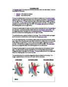
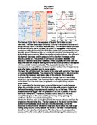
This electrical activity can be recognized as the P wave on an electrocardiogram. This electrical impulse results in contraction of the atria, pumping the blood into the ventricles.
Takes 0.045 seconds to reach AVN.
There is a delay, which allows time for the blood to flow from the atria into the ventricles. At the bottom of the right ventricle is the atrioventricular node (AV node). The atrioventricular node picks up the electrical impulse from the atria and passes it into the atrioventricular bundle (Bundle of His) which runs into the ventricles. Emerging from the A-V node, impulses enter the ventricles by way of a compact tract of conducting fibres, the bundle of His, or common bundle, which straddles the proximal portion of the intraventricular septum.
From AVN it takes over 0.120 seconds to move the next few millimetres, compared to the high speed of SAN to the AVN. This is so that it delays the impulses as you don’t want pre ventricular contractions. (Allows the atrial systole to finish before ventricular systole starts)
From there, impulses continue into the ventricles as the common bundle separates into a right branch and a left branch. These branches provide routes for rapid conduction of the impulses throughout the interventricular septum and to the endocardial surface of the free ventricular walls. The terminal elements of this conduction pathway system are the Purkinje fibres.
From Purkinje fibres, impulses penetrate to the interior of the ventricular myocardium, setting off a succession of depolarisations of sarcomeres and resulting in ventricular contraction. The depolarizing wave front progresses from endocardial to epicardial surfaces of the wall
As the electrical impulse or depolarization spreads through the ventricles, it can be recognized as the QRS complex on an electrocardiogram. This electrical activity causes contraction of the ventricles, which is also known as ventricular systole.
The spiral disposition of cardiac muscle sheets causes the spiral waves of contractions from systole for efficient basal squeeze, and the blood moves upwards, away from the spiral contractions.