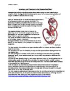
The heart is an organ, about the size of a clenched fist (pretty small for a critical role that it plays in the body), situated between the lungs and protected by the sternum (breastbone), the heart comprises of specialised cardiac muscle tissue.
The actual structure of the heart consists of 4 chambers, 2 chambers on each side, separated by the septum.
The septum ensures that neither oxygenated blood and deoxygenated blood mix together. This ensures that blood with the maximum oxygen saturation is delivered to the cells. This feature is found in a ‘double circulation’ system, where the blood goes through the heart twice, for every ‘lap’ around the body.
The top 2 chambers are the left and right atria, and these are used to receive blood from the veins. The right atrium receives blood from the superior vena cava (deoxygenated blood from the head), and the inferior vena cava (deoxygenated blood from the body).
Both venae cavae have valves to maintain a one-way flow of blood, this prevents blood back flowing due to gravity and reduced pressure in the veins. As the atria fills up with blood, the heart goes in atrail systole, where the atria contract and the blood is forced through the atrio-ventricular valves (tricuspid in the right hand side/bicuspid in the left hand side) and into the ventricles below.
The left ventricle is significantly larger than the right. This is because it does a lot more work than the right. The left ventricle is responsible for pumping the blood with enough pressure for it to go round the whole body. The thick muscular walls which line the left ventricle is there to withstand the pressure, and also, for the extra ‘pumping power’ needed.
The right ventricle however, is responsible for pumping the blood through the pulmonary artery and into the lungs, as part of the pulmonary circulation system, where the blood diffuses waste gases (carbon dioxide) out, and oxygen in.
During ventricular systole, when the ventricles contract to pump the blood, more valves come into action These are the semi-lunar valves, and these prevent the blood flowing back into the heart after going into systole.
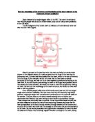
With all the blood passing through the heart, the heart itself doesn’t get any of the oxygen itself from the blood within the chambers. Instead, the heart is supplied with its own oxygen and food from the coronary arteries, which cover the entire heart. The two major coronary arteries, (the right coronary artery and left coronary artery) branch off the aorta, which then divides into several smaller arteries that goes into the cardiac muscle and supplies the heart with blood.
What also makes the structure of the heart so unique is the muscle, which makes up this pump. Cardiac muscle differs from the rest of the body because it is myogenic. This means, that the muscle naturally contracts and relaxes without any instructions from the brain. Special ‘pacemaker’ cells in the SAN (Sinoatrial Node) co-ordinate with AVN (Atrio-ventricular node) cells to create a controlled, regular cycle of contractions of the myogenic cardiac muscle. This is what makes the cardiac cycle.