The Uses of X-rays in Radiotherapy
There are two types of radiation used to treat cancer. These include Photon radiation and Particle radiation.
Photon radiation
Early radiation therapy was accomplished by x rays and gamma rays. X rays and gamma rays are essentially high energy, ionizing electromagnetic rays composed of mass less particles of energy called photons. The distinction between the two is that gamma rays originate from the decay of radioactive substances (like radium and cobalt-60), while x-rays are generated by devices that excite electrons (such as cathode ray tubes and linear accelerators). These ionizing rays are part of the electromagnetic spectrum, as are ultraviolet, visible, and infrared light, radio waves, and microwaves. They act on cells by disrupting the electrons of atoms within the molecules inside cells. These atomic changes disrupt molecules and hence disrupt cell functions, most importantly their ability to divide and make new cells.
Particle radiation
Proton rays consist of protons, which have mass and charge, rather than photons, which have neither mass nor charge. Like x-rays and gamma rays, proton rays disrupt atomic electrons in target cells. The advantage of proton rays is that they can be shaped to conform to the shape of the tumor more precisely than x-rays and gamma rays. They consequently cause less injury to surrounding tissue and fewer side effects and allow delivery of higher radiation doses to tumors without increasing damage to the surrounding tissue. Proton therapy can therefore be more effective and require fewer treatment sessions.
Neutron therapy is a second type of particle radiation. Neutron rays are very high-energy rays, composed of neutrons. They disrupt atomic nuclei rather than electrons; the likelihood of cells repairing this kind of damage is very small. Neutron therapy can also effectively treat larger tumors than conventional radiation therapy. The central parts of large tumors lack sufficient oxygen to be susceptible to damage from conventional radiation, which depends on oxygen, but neutron radiation can do its damage in the absence of oxygen, so it can kill cells in the centers of large tumors. Neutron therapy has been shown to be especially effective for the treatment of inoperable salivary gland tumors, bone cancers, and some kinds of advanced cancers of the pancreas, bladder, lung, prostate, and uterus.
Another promising type of neutron therapy, neutron capture therapy. It has the ability to deliver high doses of radiation to a very limited area. A drug that binds to tumor cells but not to other cells is chemically combined with boron and then given to the patient. The tumor is then irradiated with neutrons. When the neutrons interact with the boron atoms, the boron nuclei split, creating tiny nuclear fission events just big enough to kill one cell.
High-energy radiation kills cells by damaging their DNA and thus blocking their ability to divide and proliferate. Radiation kills normal cells about as well as cancer cells, but cells that are growing and dividing quickly (such as cancer cells, skin cells, blood cells, immune system cells, and digestive system cells) are most susceptible to radiation. Most normal cells are better able to repair radiation damage than are cancer cells. Radiation treatments are parceled into component treatments that are spaced throughout a given time interval (usually about seven weeks). Thus, cells are given a chance to repair during the time between treatments. Since the repair rate of normal cells is greater than the repair rate of cancerous cells, a smaller fraction of the radiation-damaged cancerous cells will have been repaired by the time of the next treatment. This procedure is called "fractionation" because the total radiation dose is divided into fractions. Fractionation allows greater killing of cancer cells with less ultimate damage to the surrounding normal cells.
The Use of X-ray in Imaging
Wilhelm Conrad Roentgen found that the rays could penetrate a person's hand, and the outline of bones could be seen on a chemically coated fluorescent screen behind the hand. Roentgen then replaced the screen with photographic film to make lasting pictures of the images. Today X-ray film, or radiography, studies are being replaced by digital (computerized) examinations, which may be viewed either as film or on a video screen. These improved techniques and machines have minimized radiation doses and thus potential radiation injury due to exposure from X rays. These machines include the CAT scan, MRI, Ultrasound, PECT and SPECT.
Computerized Axial Tomography
CT scan is an advanced system for medical X-ray imaging of the interior of the body. In X-ray technology tomography refers to a method for obtaining sectional views of the body that eliminate X-ray shadows of body structures before and behind the desired section. The term axial indicates a series of cross-sectional X-ray images made along a chosen body axis, and the term computerized indicates that the series of images is then combined into a single three-dimensional image by means of a computer. CAT-scan images provide much more detailed information than ordinary X-ray pictures.
A patient undergoing a CAT scan is placed on a table and introduced into the large coil of an X-ray tube that is then rotated around the desired head or body section of the patient. Electronic sensors pick up the emerging rays as a pattern of electrical impulses that are fed into a computer and integrated into a single image. This image can be displayed on a television screen and photographed, and the data can be stored on a computer disk for further reference.
Succession of slits into a narrow beam and made to strike a suitable crystal at an angle. An X-ray detector, such as an ionization chamber, is arranged to intercept the reflected beam. The angle is changed by rotating the crystal and the detector, and the intensity is measured from the response of the detector.
Magnetic resonance imaging (MRI)
Magnetic resonance imaging (MRI) produces two-dimensional visual images of internal structures in any plane using strong electromagnetic fields. In MRI, the patient is placed inside a powerful magnet that aligns the hydrogen atoms in the body tissues. A radio signal is directed to the body part being examined, temporarily disrupting this alignment. When the radio signal stops, the hydrogen atoms return to alignment, but not all at the same time, since different body tissues align at different rates. A computer measures the change in realignment and converts the data into an image in any desired plane. MRI is used to evaluate injury and disease in all the organs of the body. MRI can image, in motion, the heart, its valves, and moving blood in the veins and arteries. This technique can evaluate brain function based on blood flow. Metabolic activity in organs can be measured by MR spectroscopy.
Ultrasound
Ultrasound produces images using high-frequency sound waves. When these sound waves come into contact with structures within the body, part of the sound energy is reflected back to the body surface, where it is converted electronically into a picture. This technique is commonly used during pregnancy to determine if multiple fetuses are present, or the position and age of a fetus. An echocardiogram is an ultrasonic technique used to examine patients with congenital or acquired heart conditions, and Doppler ultrasound detects blood flow in the veins and arteries.
The field of radiology also includes nuclear medicine, in which radioisotopes are introduced into the body. The isotopes are monitored as they travel through the blood vessels to determine if specific organs are functioning properly. Special cameras measure the intensity of the radiation released by the isotope and allow visualization of parts of organs not usually seen by X rays. Radioisotopes are also used to send a therapeutic dose of radiation to a specific site to kill cancer cells.
Positron Emission Tomography
Position Emission Tomography (PET), uses an isotope that emits a radioactive particle called a positron. These isotopes attach to a variety of chemicals, such as glucose, that are found in the body's metabolism. This procedure not only detects the anatomy of an organ, but also the status of its metabolic function, such as brain activity and blood flow. It is also useful in diagnosing cancer and Alzheimer disease.
Single Photon Emission Computed Tomography
Single Photon Emission Computed Tomography (SPECT) is a recently developed technique that is just beginning to have medical applications. Like PET, SPECT tracks a radioactive isotope and a computer translates the data into an image showing details about metabolic function. SPECT is proving particularly successful in monitoring heart function.
Fluoroscopy
Fluoroscopy, another type of X-ray procedure, produces images of internal body movements on a video screen in real time while the movement is occurring. Images of the heart expanding and contracting, or intestinal motility, can be recorded on videotape or disc to be evaluated at a later time.
Conclusion
Based on the findings of this research the uses of x-ray in radiotherapy and imaging plays a vital role in the medical industry. Due to advance technology x-rays have helped to eliminate cancer cells that reproduce rapidly affecting parts of the human body. Today, an increasing number of patients have their cancers treated successfully, with few side effects such as fatigue, low blood counts, difficulty or pain swallowing, hyper pigmentation, skin itching, vomiting and nausea that previously occurred. Using radiation therapy preserves normal cells in the body. Modern technology has combined the use of three-dimensional imaging technology, computerized treatment planning and high-energy x-ray machines to make this possible.
Bibliography
http://cis.nci.nih.gov/fact/7_1.htmNokia
Appendix A
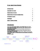
Table of Contents
Acknowledgement…………………………………………………………………………………
Introduction……………………………………………………………………………………….
Table of Contents…………………………………………………….……………………………
Literature Review
What is an X-ray?…………………………………………………………………………………..
Applications of X-rays……………………………………………………………………………..
Radiotherapy
What is Radiotherapy?……………………………………………………………………………..
The uses of x-ray in radiotherapy………………………………………………………………….
Photon Radiation…………………………………………………………………………...
Particle Radiation…………………………………………………………………………...
Neutron Therapy……………………………………………………………………………
Neutron Capture Therapy…………………………………………………………………
The uses of x-ray in imaging
Computerized Axial Tomography……………………………………………………………
Magnetic Resonance Imaging………………………………………………………………
Ultrasound…………………………………………………………………………………..
Nuclear Medicine……………………………………………………………………………
Position Emission Tomography……………………………………………………………
Single Photon Emission Computed Tomography……………………………………………
Flouroscopy…………………………………………………………………………………
Conclusion…………………………………………………………………………………………..
Bibliography…………………………………………………………………………………………
Appendices………………………………………………………………………………………….
Appendix B
Title Page
Name: Diedre Rodney
School: Clarendon College
Form: 6A
Reg no.
Centre no.
Teacher: Mr. Pryce
Territory: Jamaica