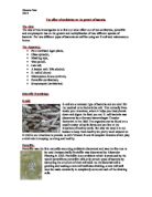
Penicillin is still used clinically although not as often as it once was due to the development of resistance by microorganisms and people’s allergic reactions. However it is still prescribed to treat syphilis, gonorrhoea, meningitis and anthrax. The success of Penicillin led to a search for other antibiotic-producing microorganisms. It was this search that led to the discovery of Streptomycin.
Streptomycin was found in 1943 from the microorganism Streptomyces griseus, in the soil environment. It was discovered by Selman Waksman who later received the Nobel Prize in 1952. It acts by limiting bacterial protein synthesis. All normal protein synthesis inhibitors affect events that occur at the ribosome and never with amino acid activation or attachment to a particular tRNA. Streptomycin acts by binding to a specific S12 protein in the 30S ribosomal subunit and causes the ribosome to misread the mRNA so that the wrong amino acids are incorporated into the polypeptide. This results in a slow bacterial growth rate and at high concentrations can lead to cell death. The stage in translation that Streptomycin inhibits is shown below:
Streptomycin is an odourless off-white powder and is bitter to taste. It is effective against gram-negative bacilli as well as many cocci. The antibiotic is also used against tubercle bacilli and is included in a combination of other drugs to treat tuberculosis.
Streptomycin is also used in humans to treat urinary tract infections, usually through intramuscular injections for seven to ten days, as it cannot be taken orally. Studies on rats indicate that the drug does not have the potential to cause cancer but side affects can include nausea, vomiting and even injury to the kidneys and nerve damage that can result in dizziness and deafness.
The human antibiotic drug is also used as a pesticide, since it was first registered as a pesticide in 1955, to control bacteria, fungi and algae. The use of Streptomycin to control fireblight on apples and pears accounts for 58% of its total use. Other significant uses are in landscape maintenance and on tobacco. The antibiotic cannot be used for aquatic uses, as it is slightly toxic to cold water and warm water species of fish.
The other two antibiotics being used in this investigation are fresh garlic and odourless garlic.
Garlic is a herb and is most commonly known for its distinctive smell and taste and its use in various dishes. Most people are unaware of the medicinal properties that the herb possesses. These properties were first recognised thousands of years ago by Egyptians, Asians, Greeks and Indians who used it in natural remedies. Louis Pasteur first proved the antibiotic properties of garlic in 1858 when he showed how it could kill bacteria in culture dishes.
The active ingredient, which is responsible for garlic’s anti-viral and anti-fungal properties, is allicin. This is also the ingredient that is responsible for the smell. A peeled garlic clove has little smell but as soon as it’s crushed, the aroma is overwhelming. This is because alliin, a precursor molecule, found within the mesophyll cells and the enzyme alliinase, which is found in the cells around the vascular bundle are physically separated within the garlic clove. When the clove is crushed, the two come into contact and immediately produce allicin and other thiosulphates.
The allicin in garlic kills bacteria by what is known as the macroeffect. This involves interfering with the cell membrane biosynthesis; preventing the production of DNA polymerises and inhibiting RNA synthesis. By doing this, the process responsible for cell replication is disrupted.
Allicin also destroys the SH groups in proteins. These groups are found in thiol enzymes in bacteria, virus and protozoa. Antibiotics tend to target a single metabolic pathway so that the bacteria are unable to use the pathway anymore. In time, however, bacteria can find an alternative route and become resistant to the antibiotic. Resistance to the allicin in garlic cannot be accomplished, simply because it affects groups found in too many enzymes for the bacteria to find different ways around it.
Since no resistance can be built up, garlic has many uses. These include the healing of many types of common illnesses such as ear, throat and mouth infections, influenza, colds, asthma and catarrh to name a few. Lower levels of heart disease found in Mediterranean and Asian countries, where larger quantities of garlic are consumed, have led scientists to believe that the antibiotic also helps to reduce cholesterol levels and reduce the tendency of blood to clot. The body’s immune system can also be improved by taking regular garlic supplements.
Despite its many benefits, people tend to avoid taking regular supplements of fresh garlic because of its lingering odour. This is why odourless garlic capsules appear to be more favourable. However, the manufacturing of odourless garlic capsules involves removing the pungent odour which means the removal of allicin as it is this that causes the smell. Therefore the active ingredient in the garlic is not present so it is no longer effective against bacteria.
Modern technology has developed a process by which the allicin content is still present and the aroma is removed. This is achieved by blending the garlic with citrus juices and oils to deodorise the valuable ingredient. Another method is to postpone the conversion of Alliin to Allicin until after the product is digested. These new processes mean that people can benefit from the natural antibiotic without being deterred by the offensive odour.
Predictions
Bacillus subtilis
Penicillin G
I predict that Penicillin G will work effectively to inhibit the growth of B.sub. Bacillus subtilis is a gram-positive bacterium and from my research I know that Penicillin G is capable of inhibiting this type of bacteria by preventing cell wall synthesis. Therefore there should be a noticeable zone of inhibition when the agar plates are removed from the incubator.
Streptomycin
Streptomycin is most effective against gram-negative bacteria and as bacillus subtilis is a gram-positive bacterium, the growth of bacteria around this antibiotic should not be inhibited. However, Streptomycin has a broader spectrum than Penicillin and could prove to be effective against Bacillus subtilis as well as E.coli, but to a smaller extent.
Fresh garlic
Garlic has been used as a natural remedy for centuries to destroy bacteria. The allicin is the active ingredient responsible for its medical properties. This is released when the garlic is crushed as the alliin in the garlic clove comes into contact with the enzyme alliinase in the vascular bundle. The many properties that garlic possesses suggest that it would be effective against Bacillus subtilis; therefore I think there will be a noticeable zone of inhibition.
Odourless garlic
Odourless garlic capsules are popular, as they do not possess the pungent lingering odour that makes the fresh herb so easily recognised. However, once the garlic has been condensed into capsules, the active ingredient allicin is lost through the smell. If this ingredient has not been somehow preserved in the capsules used in the investigation then they will not be effective in inhibiting the growth of bacillus subtilis.
Escherichia coli
Penicillin G
From my research, Penicillin G is only effective against gram-positive bacteria therefore I expect to see no inhibition of E.coli. This is because the mode of action of this antibiotic is not effective against the different structure of the gram-negative cell wall. Penicillin G can only inhibit cell wall synthesis in gram-positive bacteria.
Streptomycin
This antibiotic is very effective in destroying gram-negative bacteria by inhibiting protein synthesis. Since E.coli is a type of gram-negative bacteria, I predict that there will be a zone of inhibition surrounding Streptomycin, as the antibiotic will prevent the processes occurring at the ribosome.
Fresh garlic
I predict that the fresh garlic will be successful in inhibiting Escherichia coli. Allicin is proven to be effective against Escherichia coli. The allicin prevents cell membrane biosynthesis, inhibiting DNA polymerises and RNA synthesis, therefore disrupting the enzyme system that is responsible for cell replication.
Odourless garlic
The active ingredient, allicin, is likely to have been lost at some point when the capsules were manufactured, therefore it will not be present to inhibit the growth of E.coli. New methods to preserve the allicin in the capsules are very unlikely to have been applied to the capsules that are to be used in the investigation. Therefore, I predict that the odourless garlic capsules will not prevent the growth of Escherichia coli.
Hypotheses:
- Odourless garlic will not be effective against either of the two bacteria.
- Penicillin G will only inhibit the growth of gram-positive bacteria.
- Streptomycin will have (little or) no effect on gram-positive bacteria.
- Both gram-positive and gram-negative bacteria will be inhibited by fresh garlic.
Preliminary work
Prior to the investigation, I practised aseptic techniques and prepared plates of bacteria. This meant that I learnt the importance of flaming metal instruments and sterilising everything, as it is easy to contaminate the nutrient agar plate.
I was able to practise inoculating the plates. First I tried preparing a streak plate (as shown in the first diagram below) so that I knew how much pressure I could apply to the agar with the wire loop without penetrating it. I then tried preparing a full lawn of bacteria. At first, I spread the bacteria on the agar with the petri dish standing up the right way to get used to the technique and then tried the same technique with the wire loop but had the petri dish upside down to reduce the risk of airborne contamination. Starting at one edge of the plate, I was taught to aim to cover the entire agar as if I was colouring in, ensuring that the wire loop reached all of the edges and finished at the opposite edge (diagram 2 below). I then rotated the plate 90 and repeated this technique to make sure as much agar was covered as possible as shown by the third diagram, not forgetting to seal the dish and placing it upside down in the incubator.
After practising this technique, I had a good idea of the coverage needed in order to see zones of inhibition.
Procedure
Variables
- I will use two different bacteria. These are Bacillus subtilis and Escherichia coli.
- I will use the same batch of B.sub and E.coli throughout the investigation.
- I will use four antibiotic agents. These are Penicillin G, Streptomycin, fresh garlic and odourless garlic.
- I will use the same nutrient agar.
- I will leave all the inoculated agar plates in the incubator for the same amount of time, which will be 24 hours. If they are left any longer than this, some zones of inhibition might be too big to measure and merge into others.
- I will use the same size of filter paper discs.
- The windows will be kept closed so the conditions in which the investigation is carried out will be kept constant.
- I will keep the temperature in the incubator constant at 30 C, as this is the optimum temperature for most bacteria.
Safety
During the practical work of the investigation, the following points (taken from Microbiology and Biotechnology) should be carried out and taken into consideration at all times.
- A student who is unwell should not take part.
- Any cuts/abrasions should be covered with a clean, waterproof dressing.
- A laboratory coat should be worn to protect clothes and reduce the risk of contamination. The coat should be washed after use.
- No eating, drinking or smoking during practical work.
- Windows and doors should be kept closed to reduce airborne contamination.
- Hands should be washed with anti-bacterial soap before and after the practical.
- Benches should be wiped down with disinfectant- 1% virkon cleaner, before and after the practical.
- Report any spillage or breakage to the teacher immediately as spillages must be treated with sodium chlorate (I) and left for 10minutes before wiping with a disposable cloth.
- Avoid all hand-to-mouth operations so as not to accidentally ingest microbes.
- Tape petri dishes after inoculation and label the base clearly with name, date and nature of inoculum to avoid microorganisms escaping into the environment if the dish should be dropped and pathogens can be easily identified if contamination occurs.
- Always work near a Bunsen burner, as it is more convenient to sterilise instruments. It also creates an updraft of air, which helps to prevent microorganisms from the worker being transferred to the culture.
- Sterilise all implements before use. Metal instruments should be heated to red heat before and after every contact with microorganisms.
- Petri dish lids should not be laid on the bench, but held while the vessel is being used.
- The necks of all tubes, bottles and flash should be passed through the Bunsen flame before and after all operations.
- Never incubate microbiological cultures at more than 30 C.
- Never examine live cultures of bacteria and do not open petri dishes of bacteria once colonies have grown.
- Sterilise all media and containers by autoclaving before use. Dispose of all cultures in plastic petri dishes by placing then in an autoclavable plastic bag and autoclave at 103Kpa, 121 C for 20 minutes.
Equipment list and justification
Methods
Method 1- Preparation of sterile agar plates
- Arrange sterile petri dishes on a clean surface, unopened.
- Set up a Bunsen burner with a roaring flame on the same bench.
- The molten nutrient agar should be readily prepared in a conical flask as it is against regulations for students to prepare the sterilised nutrient media from the powdered form.
- The molten agar should be poured between 42 C and 50 C.
- Hold the flask in one hand and remove the plug, keeping it between the third and little finger.
- Flame the mouth of the flask to ensure everything is kept sterile.
- Lift a petri dish lid just enough to pour in the molten agar until it reaches about half way up the dish.
- Replace the petri dish lid quickly.
- Shake the petri dish gently to make sure the agar is distributed evenly across the bottom of the petri dish.
- Flame the neck of the flask in the Bunsen flame for 2-3 seconds.
- Repeat this method until all six petri dishes have been done.
- Discard the empty flask and the plug into disinfectant.
- Leave the plates for approximately 15 minutes to solidify.
Method 2- Inoculating the agar plates with bacteria
- Sterilise the bench with Virkon solution and wash hands.
- Set up a Bunsen burner with a roaring flame and place the petri dishes upside down on the bench.
- Flame the wire loop until it glows red.
- Still holding the wire loop near the updraft of air from the Bunsen burner, with the same hand remove the lid from the bacteria using the little finger and flame the neck of the bottle through the Bunsen flame.
- Insert the wire loop (which should be cool) into the bottle and gently stroke the bacteria.
- Flame the neck of the bottle again and replace its lid.
- Lift the base of the petri dish (the half of the petri dish containing the nutrient agar) just enough to allow the wire loop inside.
- Starting from one side, streak the surface from side to side, using the latter technique practised in my preliminary work and ensuring there are no gaps.
- Rotate the petri dish 90 and repeat the streaking technique to make sure the whole surface is covered (as shown in the diagram below).
- Replace the base of the petri dish.
- Flame the wire loop to get rid of any remaining bacteria.
- Label the petri dish with name, date and the name and batch of the bacteria used.
DIAGRAM OF FULL LAWN
Method 3- Transferring the antibiotic agents to the inoculated agar plates.
- Divide the petri dish base into four even sections with a permanent marker pen.
- Sterilise a white tile with 1% Virkon cleaner.
- Transfer the Penicillin G and the Streptomycin onto the white tile.
- To transfer the above antibiotic agents, flame the mounted needle and gently poke the antibiotic just enough for it to stick and pick it up.
- Lift the base of the petri dish and gently poke the centre of one of the four sections. The gentle motion should be enough for the antibiotic to stick to the agar, being careful not to stroke the agar, as this would spread the antibiotic agent and alter any zones of inhibition that might form.
- Flame the mounted needle.
- Label the section with the antibiotic after each transfer.
- For the transfer of the fresh garlic and the odourless garlic, several discs of filter paper are needed. These are easily obtained using a hole punch.
- For the fresh garlic, peel a garlic clove and cut it into small pieces on the white tile using a scalpel that has been flamed beforehand.
- Use a pestle and mortar to squeeze the juice out of these pieces of garlic.
- Flame the mounted needle and use it to pick up another disc of filter paper and cover it with the fresh garlic juice.
- Use the mounted needle to pick it up again and apply it to one of the sections in the same way as the Penicillin G and the Streptomycin, remembering to flame the needle again afterwards.
- For the odourless garlic, use the mounted needle to pick up another disc of filter paper and cover it in the contents of the odourless garlic capsule on the white tile.
- Use the mounted needle to pick up the piece of filter paper, lift the base of the petri dish and gently poke the centre of the agar in the remaining section.
- Remember to label each section with the antibiotic using the permanent marker pen.
- When all the antibiotic agents have been successfully transferred to the plate, seal it with two small pieces of sellotape (as shown in the diagram below). Never place the tape all around the circumference of the dish as this prevents oxygen entering the dish and creates anaerobic conditions in which harmful pathogens are able to grow.
- Place the labelled petri dish upside down in the incubator so that the bacteria are able to respire and grow.
- Leave the plates in the incubator for 24hours, after which period they can be removed and any zones of inhibition can be measured. The zone of inhibition is the distance from the disc to the outermost section around the disc where no bacterial growth is present.
DIAGRAM OF DISH WITH ANTIBIOTIC AGENTS
Method justification
The above methods are the best way, using the equipment described in the equipment list, to ensure that aseptic techniques are used throughout the investigation. The equipment chosen is to ensure the investigation is as accurate and precise as possible and to take into account the precautions outlined under the safety subheading.
Range and number
I will use two different bacteria in this investigation. These are Bacillus subtilis and Escherichia coli. The four different antibiotic agents being used are Penicillin G, Streptomycin, fresh garlic and odourless garlic.
I will prepare 6 petri dishes and then inoculate three of them with B.sub and the other three with E.coli so that I have two repeats for each bacterium, making the investigation more reliable. I will then work with someone else’s results who has worked well using aseptic techniques and has used the same batch of bacteria. Therefore I will have six results for each bacterium. This means that I will be able to compare results to observe how similar they are and recognise any anomalies more easily.
Bibliography
Microbiology and Biotechnology
Biology 1
Encarta
helios.bto.ed.ac.uk/bto/microbes.penicill.htm
www.nlm.nih.gov
Results
Bacillus subtilis (x)
Escherichia coli (y)
Analysis
Looking at the graph, it is clear that different antibiotics affected different bacteria. Penicillin G appears to be the antibiotic with the narrowest spectrum as it was most effective against Bacillus subtilis (with the largest zone of inhibition out of all the antibiotics) but had no effect on Escherichia coli. Streptomycin had an effect on both types of bacteria, as did the fresh garlic. Odourless garlic however did not affect either of the bacteria.
The value calculated for the test statistic is compared to a critical value. Referring to the statistical table on the next page, this critical value can be found. Using a 0.05 level of significance and ten degrees of freedom (which was calculated in the statistical analysis) the critical value for the t-test is 2.228. Theoretically, if the test statistic for the antibiotic is greater than this value, there is a significant different between the antibiotics agents effectiveness on the two bacteria. If the test statistic is less than the critical value then there is no significant difference, therefore the antibiotic agents had the same effect on gram-positive and gram-negative bacteria.
For Penicillin G, the test statistic was 18.040. this is a lot greater than the critical value of 2.228 so there is definitely a significant different between the antibiotics ability to inhibit gram-positive and gram-negative bacteria. The graph showing the average zones of inhibition supports this as the mean zone of inhibition for Penicillin G was 34.08 for B.sub and zero for E.coli. This range between the two bacteria was the largest out of the rest of the antibiotics suggesting that Penicillin G is a narrow spectrum antibiotic. Therefore it is expected that the test statistic would be so high above the critical value.
From the graph I can see that Streptomycin definitely had an effect on both gram-positive and gram-negative bacteria although it is clear that it is much more effective at inhibiting E.coli. The test statistic value of 6.269 is greater than the critical value, therefore there is a significant difference between the results on the two different bacteria. However, the test statistic is not as high for Streptomycin as it is for Penicillin G so there is a smaller difference between the results on B.sub and E.coli. This is because Streptomycin has a broader spectrum than Penicillin G, meaning that it is not specific to just one type of bacterium. As well as inhibiting all gram-negative bacteria, it also affects some gram-positive bacteria so in a way its discovery was more important than Penicillin G as it could be more widely used although they are good for different purposes.
The test statistic for fresh garlic is the only one that is below the critical value, meaning that there is no significant difference between the antibiotics ability to destroy gram-positive and gram-negative bacteria. The graph supports this statistic as the mean zone of inhibition for garlic on B.sub and E.coli only has a difference of 0.05mm. Antibiotics that are effective on both bacteria are hard to find, especially ones that bacteria are unable to build up resistance to, making fresh garlic an extremely valuable antibiotic.
No statistical analysis was made for odourless garlic as there were no zones of inhibition in either of the bacteria, therefore all of the results would have been 0, which is below the critical value. It is obvious looking at the results table and the graph that there is no difference between any of the results as the antibiotic has no effect on gram-positive or gram-negative bacteria.
Conclusion
In conclusion, using the results from the test statistic, my hypotheses were correct.
Penicillin G was clearly only active against gram-positive bacteria with the highest test statistic value. There was no visible zone of inhibition surrounding Penicillin G in full lawn plates of Escherichia coli. However, Penicillin G is a very effective antibiotic as it had the largest zone of inhibition on Bacillus subtilis at 34.08mm compared to the other antibiotics. This is because, as I discovered in my research, Penicillin G can destroy all gram-positive bacteria because they are susceptible to its method of attack by cell wall synthesis. The structure of the gram-positive cell wall has a thick layer of peptidoglycan and penicillin is able to prevent the cross-linking of small peptide chains in this peptidoglycan. It does not affect gram-negative bacteria as the layer of peptidoglycan is protected by a complex impermeable outer membrane which includes lipopolysaccharides, lipids and phospholipids.
This cell wall is therefore inaccessible to the Penicillin G antibiotic as it cannot penetrate the complex outer membrane to break down the concealed layer of peptidoglycan.
My hypothesis for Streptomycin stated that it would have little or no effect on gram-positive bacteria. From my scientific knowledge I knew that it would inhibit E.coli as it is a gram-negative bacterium and Streptomycin would attack its ribosome and interfere with protein synthesis by binding to a specific S12 protein in the 30S ribosomal subunit and cause the ribosome to misread the mRNA. The wrong amino acid is therefore incorporated into the polypeptide chain in the translation stage due to the bonding of the wrong codons and anti-codons. The bacterium then cannot grow to its full potential and eventually leads to cell death. The test statistic supports this hypothesis as there is a significant difference between the results. However, looking at the graph, the antibiotic also affected B.subtilis. This is because Streptomycin is also capable of inhibiting some gram-positive bacterium to a lesser extent, depending on the shape of the bacterium. Using the test statistic alone however, the original hypothesis was correct.
I predicted that both types of bacterium would be affected by fresh garlic and the test statistic proved this to be correct. This is due to fresh garlic’s many medicinal properties. The allicin in the garlic is responsible for the inhibition of bacteria as it can prevent the production of DNA polymerases and inhibits RNA synthesis. No resistance can be built up to stop this mode of action as it affects groups found in too many enzymes for the bacteria to find an alternative metabolic pathway around it, therefore it will always be effective against many gram-positive and gram-negative bacteria as the different structured cell walls do not have a significant effect on the ability of garlic to inhibit bacteria. This is reflected by the test statistic, which showed no significant difference between the zones of inhibitions on the two types of bacterium, therefore garlic is able to disrupt the cell replication process in both gram-positive and gram-negative bacteria. Despite its smell, this is why garlic is such a multi-purpose herb.
A test statistic could not be done for odourless garlic as mentioned before, as there were no zones of inhibition. Odourless garlic capsules were not effective against either bacterium because the allicin content had been lost in manufacture. The active ingredient that makes fresh garlic such a good antibiotic agent, killing bacteria by the macroeffect, is what produces the smell and so when the smell is removed from the compound, so are the anti-viral and anti-fungal properties that allicin possesses. Therefore the antibiotic agent can no longer prevent the production of DNA polymerases, inhibit RNA synthesis and disrupt the process responsible for cell replication.
This investigation has proved that different antibiotics are specific to inhibiting different bacteria. Understanding of how antibiotics combat different bacteria is essential to the future of microbiology and the fight against diseases.
Evaluation
The experimental procedures used were the most suitable for this investigation considering the environmental conditions and the time element.
There were no obvious anomalous results as such but in many cases there was a large range of inhibition zones. For example, in the case of garlic on Bacillus subtilis, the zone of inhibitions recorded varied from 20-36mm. This difference of 16mm could be the difference between the antibiotic inhibiting the growth of the bacteria effectively or not. This suggests that more agar plates would be useful to observe where the majority of the results lie, as the results gathered appeared to be spread out so much that it is not possible to regard the mean as completely accurate.
The time restriction that was unavoidable could have been a contributory factor to the varied results. It is possible that the time constraint made it difficult to carry out the procedure as accurately as possible. For example, transferring the antibiotics to the agar plate can be very time consuming and it is not certain that the antibiotic did not touch more of the inoculated agar than what was necessary.
Another possible explanation for the varied results could be the result of a slight difference in procedure of three of the results that were taken from another person at the same time. It is worth taking into consideration that I prepared six plates, three for each bacterium. To make sure there were enough results to analyse and form a conclusion, I used another persons results so that I had six results for each bacteria.
Inaccurate results could also be due to several reasons including incomplete covering of the agar to prepare a full lawn or incomplete covering of the filter paper in garlic juices. These both have the potential to alter the results.
The accuracy of the measurements taken can also be challenged. What measurement should be taken, for instance, if the zone of inhibition is clearly larger on one side of the antibiotic than the other? In this case, the largest measurement was taken but it is not impossible for this enlarged zone of inhibition to be the result of being unable to keep the antibiotic in one place when transferring it to the inoculated agar. The antibiotic could easily have been spread and this would almost definitely have affected the results. Alternatively, although not as likely, the smaller measurement could be the result of contamination as the antibiotic would not be able to inhibit foreign bacteria. This possibility is not as significant as aseptic techniques were used to ensure contamination did not occur. The most likely explanation is that the antibiotic was more concentrated in that area; there could have been more garlic absorbed on one side of the filter paper disc.
Improvements cannot easily be made to this type of procedure as the ability to carry out the procedure accurately only emerges with practice. The procedure is made difficult by the necessity to apply the antibiotic agents to the agar plate upside down to prevent cross-contamination. Minimising the sources of error could be achieved by working in isolation as there would be no people around and the risk of contamination would be reduced. The most significant improvement that could be made is probably making sure that the procedure is well practised.
If I repeated the practical work carried out in this investigation, I would increase the number of plates used so that the results obtained could be considered more reliable.
To expand this investigation, a possibility would be to compare the antibiotics ability to destroy bacteria over more than 24 hours, recording the zone of inhibition at intervals or alternatively timing how long it takes for the inhibition zone of certain bacteria to reach a certain point to investigate which antibiotics are faster at destroying bacteria. However, these procedures would be more complicated and therefore need careful observation and ideally carried out in a well-equipped laboratory to ensure that aseptic techniques are maintained.