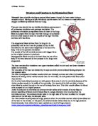
It is now that the two sides of the heart start to differ due to their functions. The ventricles at the bottom of the heart have thick muscular walls, although the left ventricle wall is thicker than the right ventricle wall. This is because the right side of the heart is the one pumping blood to the lungs. The right ventricle does not need to be as muscular because the lungs are close to the heart and not as much pressure is needed to push the blood there - if there was as much then the capillaries in the surrouding tissue of the lungs would burst and drown the alveoli in blood. The left ventricle has a very thick muscular wall because it pushes blood around the body, and so needs a high pressure to move blood in arteries in the furthest regions of the body.
When the ventricles are full, they then contract at the same time, starting from the bottom, pushing the blood upwards, through the semi-lunar valves - one on each side of the septrum. The right ventricle pumps blood into the pulmonary artery to the lungs, the left into the aorta which swells, then branches off to various parts of the body. This is called ventricular systole. When there is no blood left in the ventricles and therefore little pressure, and a high pressure in the arteries, the semi-lunar valves in the middle of the heart close to prevent backflow of blood (this makes a sound which can be heard by a stethoscope as the second thump of a heartbeat - 'Dub').
After the ventricles have contracted, the final phase of a heartbeat occurs, called diastole. This is timespan of about 0.2 seconds in which all the muscles in the heart are relaxed, allowing blood to collect in the arteries before the whole process re-occurs. This process will take approximately 0.6 seconds or 70 beats per minute, which of course varies depending on the level of activity.
This leads on to control of the heartbeat. The heart is myogenic, which means a heartbeat is initiated by the heart itself rather than by a nevous impulse from the brain, meaning that it can work without nervous delay, like a reflex action. A heartbeat is initiated by special cardiac muscle cells known as the Sino-Atrial node (or SA node), which is located in the wall of the right ventricle near the entry of the vena cavae. This sends an electrical wave of excitation spreading across both atria meaning they contract at the same time and push blood into the ventricles. The same wave of excitation will then reach and stimulate the Atrio-Ventricular node (or AV node) which causes ventricular contraction. However, blood from the ventricles needs to be pushed upwards to allow it to flow more easily into the arteries. Therefore, the new wave of excitation initiated by the AV node travels down special fibres in the septrum (mentioned before in aiding heartbeat) called Purkinje fibres, which collectively make up an insulating electrical conducting passage called the 'Bundle of His'. When it reaches the bottom of the heart, called the apex, the same wave of excitation then spreads upwards across the ventricle walls causing them to contract from the bottom up and push the blood towards the arteries. This process is called the cardiac cycle.
In conclusion, the size of the muscular walls in the heart, the existance and positioning of valves and the conduction of electrical waves of excitation make the heart perfect for the job as a pump of blood around the body.