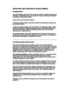
Blood Vessels
Arteries are the largest blood vessels, followed by veins and then capillaries.
Arteries
- Three Layers, they are thick and elastic- Connective Tissue, a thick muscular layer and an endothelial layer. Strength required to support pressure
- Blood is pumped away from the heart
- Blood contains oxygen (except in the case of the pulmonary artery)
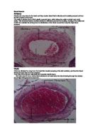
Veins
- Three Layers, they are thinner and less muscular.
- Valves prevent backflow of blood
Capillaries
- Single-celled layer, suitable for gas and nutrient exchange.
The heart walls are made of three layers:
Endocardium- the innermost layer. It is made of epithelial tissue and forms the lining of the entire circulatory system.
Myocardium- the middle layer. This layer is the thickest of the three and is made up of cardiac muscle.
Epicardium- the outer layer. The epicardium is the thin, external membrane.
.
Thrombocytes or Platelets
The main function of platelets, or thrombocytes, is to stop the loss of blood from wounds. They also can release chemicals which cause clots to form in the blood that plug up narrowed blood vessels.
Leukocytes, or white cells
Leukocytes, or white cells, are responsible for the defence of the organism. In the blood, they are much less numerous than red cells. Lymphocytes are a major part of the immune system. Other white cells called granulocytes and macrophages protect our body from infection by surrounding and engulfing the infection. They also make room for new red blood cells by getting rid of the old ones.
Erythrocytes or Red blood cells
The erythrocytes are the most numerous blood cells. The red cells are rich in haemoglobin, a protein able to bind in a faint manner to oxygen. Hence, these cells are responsible for providing oxygen to tissues and partly for recovering carbon dioxide produced as waste.
Plasma
Plasma is a clear liquid protein and salt solution which carries the red cells, white cells, and platelets about 55% of the total blood volume is made up of plasma, about 95% of it is made of water
Stroke volume is the volume of blood that is pumped out of the heart by each ventricle during one contraction
Cardiac output is the amount of blood ejected from the left ventricle in one minute you work this out by:
Cardiac output = Stroke volume x heart rate
Respiratory System
The primary function of the respiratory system is to supply the blood with oxygen in order for the blood to deliver oxygen to all parts of the body. The respiratory system does this through breathing. When we breathe, we inhale oxygen and exhale carbon dioxide. This exchange of gases is the respiratory system's means of getting oxygen to the blood.
Respiration is achieved through the mouth, nose, trachea, lungs, and diaphragm. Oxygen enters the respiratory system through the mouth and the nose. The oxygen then passes through the larynx (where speech sounds are produced) and the trachea which is a tube that enters the chest cavity. In the chest cavity, the trachea splits into two smaller tubes called the bronchi. Each bronchus then divides again forming the bronchial tubes. The bronchial tubes lead directly into the lungs where they divide into many smaller tubes which connect to tiny sacs called alveoli. The average adult's lungs contain about 600 million of these spongy, air-filled sacs that are surrounded by capillaries. The inhaled oxygen passes into the alveoli and then diffuses through the capillaries into the arterial blood. Meanwhile, the waste-rich blood from the veins releases its carbon dioxide into the alveoli. The carbon dioxide follows the same path out of the lungs when you exhale.
The main part of the respiratory system is gaseous exchange. In aerobic respiration, Oxygen must enter our blood and Carbon Dioxide must leave the blood through our lungs.
The respiratory system consists of the lungs, trachea, bronchi, bronchioles, Diaphragm and the alveoli.
Trachea-
The trachea consists of18 rings of cartilage which is lined by a mucous lining. This helps open and protect the trachea.
Bronchioles and bronchi-
The trachea divides into two bronchi the right bronchus goes into the right lung and the left bronchus goes into the left lung. The bronchioles enable the air to pass into the alveoli, where diffusion takes place.
Alveoli-
Alveoli are the tiny air filled sacs which are responsible for gaseous exchange between the lungs and the blood. There are millions of Alveoli in our lungs and they provide a massive surface area for the exchange to happen.
Diaphragm-
The diaphragm is what controls our breathing. It moves down and sucks air in to the lungs. Hic cups are caused when the diaphragm becomes out of rhythm and pushes out little shoots of air.
To test cardiovascular and respiratory levels I got a class mate to perform the Bruce treadmill test, which is a test to monitor the development of an athlete’s general endurance. It works by some one running on a treadmill for a set amount of time and as the time goes on the speed and slop increases. We set up 10 stages each stage lasts for three minutes and the slop goes up two stages at the start of each stage, and the speed goes up two stages each time until it reaches twelve. Here are the results in a table and the results is what the athlete was saying for him self:
You can see by my findings that the short term effects of the Bruce treadmill are sweat, heavy breathing, increased heart rate, muscle burn and slight pain.
This individual may not have stretched off properly before the test that is why he got pain in his hamstrings.
His heavy breathing could have been because as the levels grew faster and the incline increased an even bigger need of oxygen was needed for the working muscles. This may have lead to the muscle burn this individual has suffered due to lack of oxygen present to fight lactic acid.
If this person where to continue this exercise for the long term I would expect that this person’s cardiovascular endurance would increase and that the individual’s heart rate would decrease along with blood pressure. Hypertrophy would increase causing muscle to grow and reduce the amount of fat in this person’s body.
Indoor rowing test
This test is 2000 meters on level 5 on the rowing machines
In the 2000 metre rowing test short term effects are red faced, muscle burn and aching, sweat, heavy breathing and increased heart rate. His heavy breathing could have been because the individual may have been struggling for oxygen this is evident because the person began to go red in his face due to a lack of oxygen. Also the muscle burning would support my idea because due to lack of oxygen the muscles where not allowed to get rid of the lactic acid build up.
If the individual where to continue doing this exercise for the long term I would expect that this person’s cardiovascular endurance would increase and that the individual’s heart rate would decrease along with blood pressure. Hypertrophy would increase causing muscle to grow and reduce the amount of fat in this person’s body.
Jonathan Taylor
Btech national diploma
Sandro Sandri
Body in action Task 2
Yr 1
BLM